Hospital Tutorial Write-ups

Medical notes record the data you have gathered and your medical decision-making in a concise, well-organized, and standard way. These notes communicate your findings with other members of the team and serve as legal documentation of the care that has been provided. In FCM, your hospital tutorial write-ups will give you practice in writing comprehensive notes, like those you’ll write in third year for new patients.

Almost all health systems now use electronic health records for documentation. We teach you to write notes from scratch because the structure and your clinical reasoning should be very similar in a written and in an electronic note. Written notes will build good habits and allow your mentors to provide you with better feedback. Plus, most of our first year students don’t have access to the EHR at their clinical sites, and even if you did, we want you to learn to collect information from your patient yourself rather than use information collected by others

Your FCM write-ups should have the following structure – more details on each section are included below.

Section Content of this section
Identifying Information & Chief Concern Name, age
Known medical problems highly relevant to the chief concern (< 4)
Chief concern and duration of symptoms
History of Present illness Background: Health at the time of symptom onset and details of any chronic illness directly related to the chief concern
Details of the presenting problem beginning at symptom onset & proceeding sequentially
Predisposing conditions & risk factors
Pertinent negatives
Optional: Hospital course or evaluation to date
Past Medical History All active medical problems and any other problems relevant to evaluation or ongoing management
Summarize for each major, active problem: diagnosis, current treatment, control, and complications
Medications & Allergies Prescribed medications and doses
Non-prescription medications and complementary therapies
Drug allergies and the type of reaction
Health-Related Behaviors Substance use: Tobacco, alcohol, recreational drugs

Diet & exercise

Sexual history

Preventive Health

Family Medical History Family history of illnesses and causes of death affecting first degree relatives
Social History Full social history, including influences on your patient’s health and health care: living situation, social support, occupation and avocation, any financial or other concerns, sexual orientation (if not included in sexual history), gender identity & assigned sex at birth, cultural identity
Review of Systems List all systems and note the presence or absence of each symptom you asked about. For organ system(s) discussed in HPI, write “see HPI”
Provide details of positive responses to ROS questions
Physical Exam General appearance and vital signs
Document each organ system in order and report all exam findings, both normal and abnormal
Labs and Imaging Not part of the FCM write-up. Should be reported after the physical exam in later notes.
Summary Statement Restate the ID & summarize the key features from the history & physical exam
Branching Diagram or Diagnosis Matrix A visual representation of your clinical reasoning process. Not included in Term 3 or later write-ups.
Assessment Format determined by clinical context
Plan Includes diagnostic and treatment plan, monitoring/follow-up and patient education

Identifying data & chief concern

Purpose: Sets the stage and gives a brief synopsis of the patient’s major problem

Format:

Identify the patient by name and age. You can include gender if it has been confirmed or sex if relevant to the CC.

Include no more than 4 medical problems (sometimes there are zero) that are highly relevant to the chief concern. List only the diagnoses here & elaborate on them in the HPI or PMH.

Report the chief concern and duration of symptoms

History of present illness

HPI Background: Health at the time of symptom onset and details of any chronic illness directly related to the chief concern.
Details of the presenting problem beginning at symptom onset & proceeding sequentially.
Predisposing conditions & risk factors (‘pertinent positives’)
Pertinent negatives
Optional: Hospital course or evaluation to date

Purpose: Provides a complete account of the presenting problem. Includes any information from the past medical history, family history and social history that is directly related to the presenting problem.

Format: One framework commonly used to structure the HPI is:

Background

Characterize the patient’s health at the time current symptoms began. If the symptoms are related to a chronic illness, give a brief summary of the illness, including when it was diagnosed, treatment, complications, and how well it is controlled.

Example 1. MR was in his usual state of good health until the day before admission when …

Example 2. ST has a history of coronary artery disease and had a myocardial infarction 6 months ago.  He has had stable chest pain with exertion since that time, until the morning of admission when…

Details of the presenting problem

This is an organized and edited version of the patient’s narrative, beginning at the onset of symptoms, and proceeding sequentially to the time of presentation.  In FCM, your write-ups should be written as though you were present at the time of admission. Focus on the initial symptoms and presentation, rather than what has happened since hospitalization. This will allow you to practice clinical reasoning more effectively.

Predisposing conditions and risk factors 

These are sometimes called ‘pertinent positives’ – the past medical history, health related behaviors, family history, or review of symptoms that is directly related to the presenting problem.  For example, a family history of liver problems is pertinent and should be included in the HPI of a patient who presents with jaundice. The same family history would NOT be pertinent to a suspected UTI, so would NOT be included in the HPI.

Pertinent negatives

Pertinent negatives are the absence of symptoms from the organ system(s) involved and  negative information from the rest of the history that directly affects your assessment of the patient’s problem.  For example, “No family history of liver disease” is a pertinent negative in a patient presenting with jaundice.

Optional – hospital course or evaluation to date: 

Here you can summarize what has occurred since the admission – significant events, testing, and how the patient is doing now. Since you will be getting this information from the patient rather than the medical record, you will probably not have all of the details. Save this information until the end of the HPI and practice constructing the first paragraphs as though YOU were assessing the patient’s presenting concern, rather than relying on information about what other doctors have said or done.

Past medical history

Purpose: A comprehensive summary of all medical and surgical problems, both current and resolved.

Format: Use a bulleted list with additional details and explanations when appropriate.

    • Major childhood illnesses
    • Medical problems, including psychiatric illnesses. You can simply list problems that have resolved. For each ongoing problem, outline:
      • Onset – date and presenting symptoms or testing if known
      • Treatment and current control
      • Complications
    • Surgeries (type of surgery and date)
    • Traumas
    • Obstetrical history, if applicable

Medications & allergies

Purpose: Document medications taken at home, to ensure appropriate therapy is being given and drug interactions are minimized.  In the EHR, medications are documented in a separate area and should be updated at each outpatient visit. In the hospital, electronic chart notes may include the list of medications that are being administered based on orders and pharmacy records.

Format:

  • List all prescribed medications by generic name. Include doses if possible
  • List over the counter medications
  • List complementary/alternative medicines
  • List medication allergies including the reaction

Health Related Behaviors

Format: May be written as a bulleted list or in sentence format in FCM notes.  In an EHR, this information is often collected in a separate field. It includes:

  • Diet and exercise
  • Tobacco (in pack-years, current amount, and stop date if applicable)
  • Alcohol (past and current amount, stop date if applicable)
  • Drugs (past and current amount, stop date if applicable)
  • Sexual history (primarily sexual behavior; sexual orientation and gender identity may be included in social history)
  • Preventive health can be included but is more applicable in the clinic. Here you can note immunizations, appropriate screening (Pap smears, colonoscopy, etc.), safety

Family history

Format: List first degree relatives (grandparents, parents, siblings and children) and major diseases each was affected by, including coronary artery disease, cancer, diabetes, alcoholism, genetic diseases, or major psychiatric illness.  You may also use a genogram.

Social History This may include influences on your patient’s health and health care: living situation, social support, occupation and avocation, any financial or other concerns, sexual orientation (if not included in sexual history), gender identity & assigned sex at birth, cultural identity

Length: ~ ½ page

Purpose: To understand the impact of education, culture, religion, income and social support on the patient’s health and health care.

Format: Written in paragraph form and includes at least some of the following:

    • Occupation and current employment
    • Upbringing, education, and family relationships
    • Current living situation and sources of support
    • Financial and insurance issues
    • Stresses, satisfaction, interests
    • Religious/spiritual support
    • Cultural identity
    • Sexual orientation
    • Gender identity
Review of Systems List all systems and note the presence or absence of each symptom you asked about. For organ system(s) discussed in HPI, write “see HPI”
Provide details of positive responses to ROS questions

Length: ~ 1/2 page

Purpose: Identify additional symptoms that require attention or evaluation.  If you uncover symptoms in this section of the interview that could be related to your differential diagnosis, you report them in the HPI rather than in this section of the writeup. Remember that diagnoses belong in the HPI or PMH; symptoms belong in the ROS.

Format:

List all the review of symptoms for each system not discussed in HPI—for organ systems relevant to the presenting complaint, write “see HPI”

Provide details of positive responses to ROS questions

Physical Exam General appearance and vital signs
Document each organ system in order and report all exam findings, both normal and abnormal.

Length: 1-2 pages

Purpose: to describe your exam findings, highlighting those that support or argue against items on your differential diagnosis.

Format:

Begin with one sentence description of general appearance and comfort level

List vital signs: Blood pressure, pulse, respiratory rate, temperature, oxygen saturation (if known)

List in outline form a system-by-system description:

    • Skin
    • Head, ears, eyes, nose, throat (HEENT)
    • Chest/pulmonary
    • Cardiovascular
    • Abdomen
    • Genital/pelvic/rectal (omitted in FCM)
    • Extremities/musculoskeletal
    • Neurologic
    • Skin

Be complete: describe what you found, both positive & negative findings. Do not just write “normal.” We want you to practice the language you would use to document findings important to your differential. You can shorten your writeups later.

Do not interpret findings. Document only what you see, hear or feel

Labs and Imaging Results of testing are documented after the physical exam in Clerkships
Summary Statement Restate the ID & summarize the key features from the history & physical exam

Length: 1-2 sentences

Purpose: Synthesize the important history and exam findings, to frame the clinical problem and to lead your listener to your assessment. This is not simply a restatement of the ID chief concern as you add key features identified in your H&P.  It is similar to the mental ‘problem representation’ created by experienced clinicians as they perform an H&P.

Branching Diagram or Diagnosis Matrix Visual representation of your clinical reasoning process, included in Term 1 write-ups
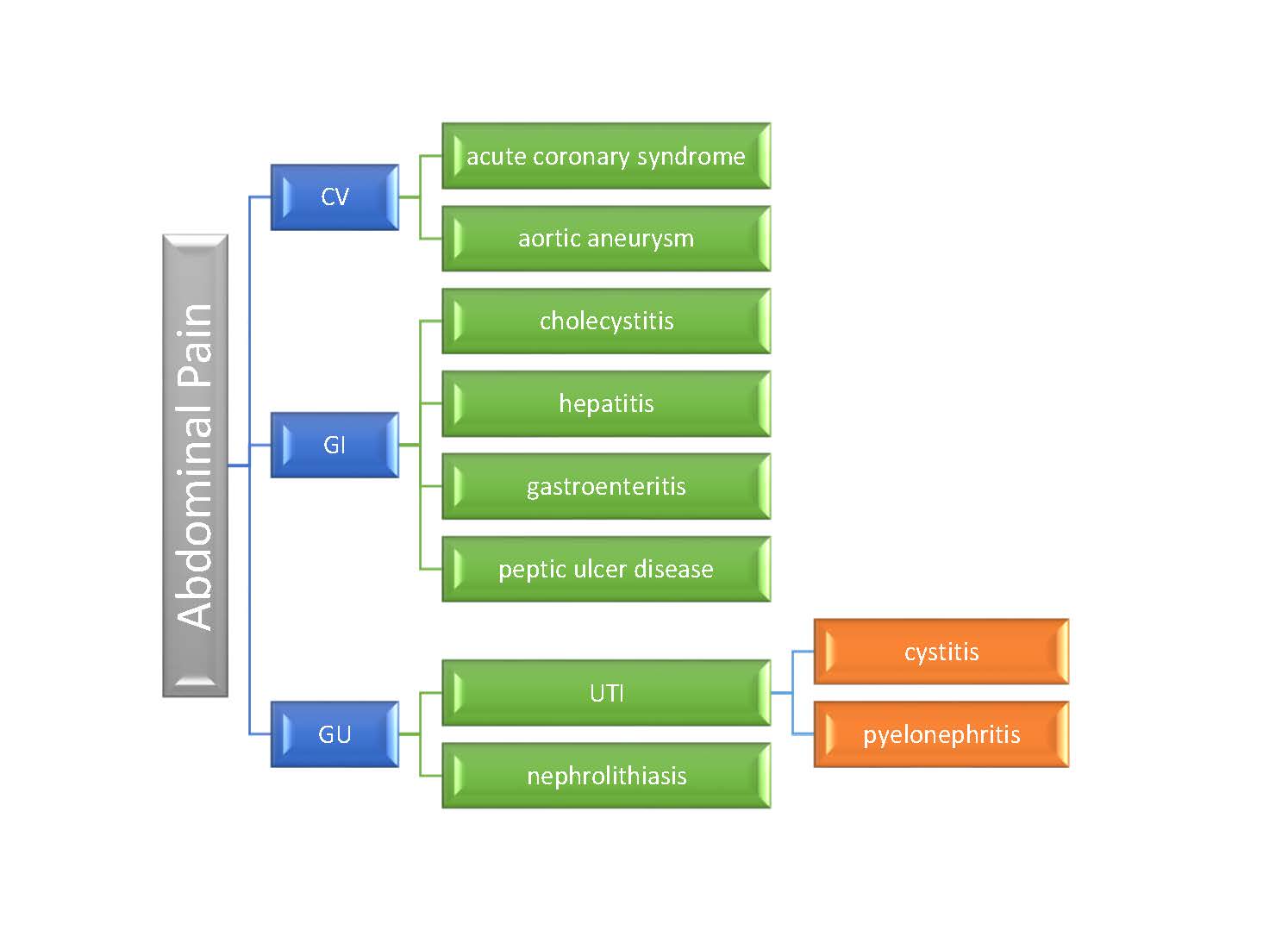
Branching diagram

Purpose: A branching diagram is an educational tool that will help you develop and organize a complete differential diagnosis. You will not see a branching diagram in real notes, but you will see your teachers use a similar structure to outline their approach to common concerns.

Format:

Begin with a symptom, physical exam finding, or other interesting feature of the case

Link possible causes to the primary symptom/finding/feature in an organized manner.  Templates are available on Canvas.

Diagnosis Matrix

Purpose: Another tool for organizing and elaborating on your differential diagnosis. This matrix format allows you to compare the history, risk factor & exam finding of your leading possibilities. You will include a matrix in your Term 2 writeups.  A template is available on Canvas.

Format:

Begin with a differential of the 3-5 most likely or “can’t miss” causes of the patient’s presenting problem

Record the typical or common findings in each section of the matrix, and bold or highlight those present in your patient.

BILATERAL LEG EDEMA

History of Present Illness

Predisposing conditions & Risk Factors

Physical Exam Features

CHF

Edema

Dyspnea, orthopnea, PND

Weight gain

Decreased exercise tolerance

Coronary artery disease

Severe lung disease

Cardiac risk factors

Elevated JVP

Enlarged or displaced PMI

S3

Crackles

Hepatojugular reflux

Cirrhosis

Edema

Increased abdominal girth

Weight gain

Jaundice

Alcohol Use Disorder

Viral hepatitis

Injection drug use

FHx of liver disease

Normal JVP

Ascites

Spider angiomas

Jaundice

Splenomegaly

Venous Stasis

Edema

Skin discoloration or ulceration

Prior DVT

Obesity

Hemosiderin deposition in skin

Ulcerations

Normal cardiac exam

Assessment Format determined by clinical context

Purpose: Explicitly state your clinical reasoning after you have read about your patient’s chief concern or medical problem.

Format for a new problem:

Begin with a differential diagnosis of 3-5 possible causes of the patient’s problem.

Discuss the most likely cause of the patient’s problem. Link the features of your patient’s history and physical that either support or argue against this diagnosis. It is rare that a disease on the differential is completely “ruled in” or “ruled out” based on the H&P.  Use terms like “most likely because” or “very unlikely given” rather than the more definitive “rules in” or “rules out”.

Next discuss the other 2-4 other diagnoses you are considering (based on your branching diagram or matrix). Again, link the features of your patient’s history and physical either support or make each diagnosis less likely

Format for an exacerbation of a chronic problem:

Your assessment would address the most likely reasons for the exacerbation, as suggested by your patient’s history and exam findings.

Plan Include in Term 2 Spring Quarter & Term 3 writeups

Purpose: To outline your next steps in addressing your patient’s clinical problem(s).

Format: The plan is usually presented as a bulleted list, and may include interventions in these categories:

Diagnostic Plan Lab tests
Imaging
Consultation with specialists
Treatment (Therapeutic Plan) Behavior change
Medications
Counseling
Referral to another provider (e.g. physical therapy)
Monitoring and follow-up Repeat laboratory tests to monitor response to treatment
Routine screening tests
Primary care clinic follow-up
Education Education about diagnoses done by you
Referral to other providers for additional teaching, e.g. diabetes educator, pharmacist

Common Problems with FCM Write-ups

If your patient has a known diagnosis and is admitted for routine therapy

If your patient has a known diagnosis with no new symptoms, for example a patient with cancer admitted for chemotherapy, there is still much you can learn from an interview and write-up.  In your write-up, you can approach the case as a new referral – you are documenting the details of their illness even though the diagnosis is already known. Report on the initial presentation and progression of the illness, how it was diagnosed, treatment to date, the impact on patient and family, and what your patient is hoping for.

If two equally important problems were responsible for hospital admission

You can take either of two approaches

  • combine the two problems in the HPI and record all symptoms chronologically OR
  • separate the two problems in the HPI, first providing a complete history for the first problem and then a complete history of the second.

The chronology is unclear

Approach the patient from the standpoint of their history at the time of admission, when they presented for care. Your history is then presented in a chronology organized on the basis of time “prior to admission” (PTA). You should describe all events using consistent time points, like hours/days/months/years prior to admission.

The physical exam shows up in the HPI

Do not put physical exam findings from your examination into the history portion of the database, or historical information into the write up of the physical exam.

Nonstandard abbreviations are used 

Many abbreviations are used differently in different specialties. In addition, abbreviations are often overused – – “the pt is STH prbm c SOB…” translates to “the patient is said to have a problem with shortness of breath” and is inferior to “the patient became dyspneic…”. When in doubt, spell out the word. Unfamiliar abbreviations will only bewilder your reader, and many are not allowed because of patient safety issues.

The Physical Exam and Review of Systems are mixed up

The ROS is part of the “subjective” portion of the medical database, along with the HPI and Past Medical History. This subjective information is given by the patient, or family or caregivers. The PE is part of the “objective” portion, reflecting your own findings and observations. Next year, the ‘objective’ section will also include labs and other testing, after the PE. Never mix the “subjective” and “objective” portions of the database

Recording a “diagnosis” instead of a “finding” in your physical examination

In the physical exam section, you should describe your findings in detail. Diagnoses should be reserved for your assessment and plan.  Instead of writing “findings consistent with RLL pneumonia” you should write “Chest: symmetric excursion, non-tender to palpation; dullness to percussion right lower lung field; bronchial breath sounds with occasional mid-inspiration crackles right lower lung field on auscultation.”

Term 1 Sample writeup – acute problem

FCM Sample Write-Up #1
Term 1
Sample A: Acute Problem


ID/CC

Mr. Y. is a 56 year-old man with a history of hypertension and peptic ulcer disease who presents with 5 days of diarrhea and right lower quadrant pain.


HPI

Mr. Y was in his usual state of good health until 5 days prior to admission while on a road trip with his son in Colorado. He developed diarrhea, described as loose, somewhat watery occurring two to three times a day. The volume of the stool was not more than normal. He also had nausea but no emesis, and was able to eat/drink normally.  The stool was brown without melena or hematochezia.

At the same time, Mr. Y. also developed a dull, steady, and fairly mild pain in his right lower quadrant; “2 or 3” out of 10. If he pressed on the area, the pain became sharp and more intense. The pain did not radiate and there were no aggravating or alleviating factors. He had no jaundice, odynophagia, change in appetite, dysphagia or heartburn.

Three days prior to admission, Mr. Y.’s symptoms had not improved and he developed intermittent emesis. He developed subjective fevers and sweats. His nausea started to keep him up at night and his appetite decreased significantly. He tried taking acetaminophen for his symptoms but this did not provide any relief.

Mr. Y. returned to Seattle on the day of admission. Having not improved, he saw his family practice doctor who noted an elevated WBC on his blood work and instructed Mr. Y. to go to the ER.

While in Colorado, Mr. Y. stayed with family members who were sick with influenza. He also ate a “questionable” pork sandwich at a football game the night prior to the onset of his symptoms. His symptoms did not feel similar to those he experienced with his previous peptic ulcer disease in the 2000s, and Mr. Y. has never experienced anything like this before. Mr. Y. attributes his current symptoms to food poisoning from the pork sandwich in Colorado, but is also concerned that his appendix might be ‘acting up’.

Hospital Course: Mr. Y. had a CT scan in the ER which showed a partially ruptured appendix and he was admitted. Mr. Y. has been in the hospital for 2 days at the time of this interview. Upon arriving at the hospital, Mr. Y. had a surgical consultation for appendicitis. Surgery was not performed and Mr. Y. was administered IV antibiotics and put on bowel rest. His diarrhea, pain, and nausea all began to resolve within 12 hours of the onset of treatment. At this time, Mr. Y. has few symptoms and is feeling much better.


PMH

Major Childhood Illnesses

  • Chickenpox -age 11

Medical Problems

  • Hypertension, diagnosed 2013
    • Diagnosed with BP approximately 190/110; currently on medications with average BP approximately 160/85
    • Patient is unaware of any secondary problems/end-organ injury related to his hypertension
  • Hypercholesterolemia, diagnosed 2013
    • Currently controlled with simvastatin
    • Most recent total cholesterol checked 3 months ago: 240
  • Degenerative Disk Disease, diagnosed 2010
    • Currently controlled with chiropractic manipulation; no current weakness or numbness
  • Duodenal Peptic Ulcer Disease, early 2000s
    • Treated with antibiotics, no recurrences since

Surgeries/Trauma

  • Fingertip amputation repair- 2001

Psychiatric History

  • No history of depression or mental illness

Medications

  • Amlodipine/Benazepril, 2.5/10 ; 1 capsule by mouth every day
  • Simvastatin, 40mg by mouth every night
  • Complementary/Alternative Medication: None


Allergies: No Known Drug Allergies

Health Related Behaviors:

  • Tobacco:  Never
  • Alcohol:  5 drinks/week
  • Recreational Drugs:  None
  • Travel, Exposures: Colorado road trip (see HPI); significant travel to Asia and Europe within the past 20 years, none in the past 3 years.
  • Personal safety habits: No firearms, uses seatbelts
  • Sexual history: Female sexual partners; monogamous for 30 years; no STD testing
  • PCP is family practice doctor in Issaquah
  • Regularly visits a Chiropractor
  • Visits a Dentist about once every two years
  • Diet:  Frequently eats out as he travels a lot
  • Exercise:  No regular exercise program


FHx:

  • MGF – prostate CA; MGM – hyperlipidemia; paternal grandparents – unknown
  • Father – MI x 2 in his 50s, died @ 82, CVA
  • Mother – alive and well @ 84
  • Three adult children – alive and well


SH:

Mr. Y. is a pilot for Alaska Airlines. Recently, his pilot’s license has been suspended due to his uncontrolled hypertension. Mr. Y. seems to be handling this well, using his free time to travel to see his sons and spend more time with his wife. He reported feeling excited by the fact that this situation may lead to an early retirement. Mr. Y., whose gender identity is male, has a close relationship with his family, and relies on them for support during stressful situations. His youngest son has just left home for college, and he and his wife are adjusting well to being empty-nesters in their home in Issaquah. He played football and lacrosse when he was younger, and enjoys watching his youngest son play lacrosse on a club team. Mr. Y. has health insurance and finances are not a significant source of stress. He is a Christian, but he reports this does not play a large role in his life.


ROS

General: see HPI

Derm: No rashes, pruritus, changing moles, lumps, lesions

HEENT: no headaches or trauma;

  • Eyes: no diplopia, wears reading glasses, no change in vision, eye pain or inflammation
  • Ears: no difficulty hearing, tinnitus, vertigo or pain
  • Nose: nasal stuffiness/obstruction, no nose bleeds or sinusitis
  • Mouth: no sores, sore throat or dentures


Respiratory:
no dyspnea, pleuritic pain, cough, sputum, wheezing, asthma, hemoptysis, cyanosis, snoring, apnea, history of TB exposure, PPD

Cardiovascular: positive for HTN(see PMH), no chest pain, angina, dyspnea on exertion, paroxysmal nocturnal dyspnea, orthopnea, peripheral edema, history of murmur, palpitations, claudication, leg cramps, history of DVT

Gastrointestinal: see HPI

Genitourinary: No dysuria, nocturia, hematuria, frequency, urgency, hesitancy, urinary incontinence, urethral discharge, sores, testicular pain or swelling

Musculoskeletal: Positive for intermittent back pain, no other joint pain, swelling stiffness, or deformity; no muscle aches or locking of joints

Neurological: no dizziness, involuntary movements, syncope, loss of coordination, motor weakness or paralysis, memory changes, speech changes, seizures, paresthesias

Psychiatric: no sadness, sleep disturbance, crying spells, anorexia or hyperphagia, anhedonia, suicidal/homicidal ideation, loss of libido, anxiety, history of eating disorders, hallucination, delusions, behavioral changes

Hematologic: no anemia, easy bruising or heavy bleeding

Endocrine: No polyuria, polydipsia, head/cold tolerance

Functional status: Able to complete all ADLS without impairment (bathing, ambulating, toileting, transfer, eating, dressing). Able to complete all instrumental ADLs without impairment (shopping, cooking, mode of transportation, telephone use, laundry, housekeeping, responsibility for meds/finances).


PE

General: Mr. Y. is well-appearing, responds to questions easily and moves without difficulty, with no signs of acute distress.

Vital Signs:  BP 160/90; Pulse 60; Resp 16

Skin: Warm, dry, no rashes. Well-healed surgical scar on right forearm. Nails normal without clubbing, cyanosis, or lesions.

HEENT:

Head:  Normocephalic. Face, scalp and skull without lesions or tenderness.

Eyes: Vision 20/40 in each eye. Conjunctivae without injection, sclera anicteric. Corneal light reflex symmetrical. PERRLA. Red reflex present bilaterally. Disc margins not appreciated, retinal vessels normal in appearance and configuration. Fundi clear without hemorrhage or exudate bilaterally.

Ears:     Left ear canal with small dark lesion, right ear canal without lesions or discharge. Tympanic membranes gray-white in color without bulging or erythema.

Nose: External nose without lesions or asymmetry. Nasal mucosa pink bilaterally without lesions, septum deviated to the left, inferior turbinates visualized bilaterally without lesions or exudates.

Mouth/Throat: Mucosa pink without lesions. Left lower molar dental carries. Uvula midline. Tonsils and posterior pharynx without erythema or exudate.

Neck/Thyroid: No palpable cervical lymph nodes. Thyroid normal in size and consistency, non-tender.

Chest/Lungs: Breathing symmetrical without use of accessory muscles. No tenderness on percussion of spine or CVAs. Lung fields resonant to percussion. Lungs with normal bronchovesicular breath sounds without wheezes or rales.

Cardiovascular:

Regular rate and rhythm, S1 single, S2 physiologic split. 

Pulse DP PT R C
Right 1+ 1+ 2+ 2+
Left 1+ 1+ 2+ 2+

Abdomen: Normal bowel sounds. Tenderness to deep palpation of the right lower quadrant at McBurney’s point without guarding or rebound. No tenderness on deep palpation of the other quadrants. Liver 12 cm on percussion. Spleen not palpable. No masses.

Musculoskeletal:  Normal range of motion of all four extremities. No lower extremity edema.

Genital:  Omitted

Rectal:  Omitted

Neurologic:

Mental Status: Alert, oriented to person, place, and time. Speech fluent, articulate, and appropriate.

CN II: Tested (vision, pupillary light reflex) and Intact

CN III, IV, and VI: Tested (extraocular eye movements) and Intact CN V: Tested (masseter muscles) and Intact

CN VII: Tested (facial contortion) and Intact CN VIII: Tested (finger rubbing) and Intact CN IX and X: Tested (phonation) and Intact

CN XI: Tested (shrug shoulders/turn) and Intact CN XII: Tested (protrude tongue) and Intact

Motor: Strength 5/5 throughout all muscle groups. Normal, fluid movements observed without tremor.

Sensation:  Able to feel sharp and dull appropriately on hands and feet bilaterally.

Romberg negative. Reflexes: Biceps, triceps, knees, and ankles 1+ bilaterally. Toes downgoing bilaterally.

Cerebellum: Finger-nose-finger test smooth bilaterally; heel-shin smooth bilaterally. Gait normal.

Summary

Mr. Y. is a 56 year old man who presents with fever, diarrhea and RLQ abdominal pain of 5 days duration and has a physical exam remarkable for lower right quadrant abdominal tenderness on deep palpation without guarding or rebound pain.

Diagram of abdominal pain. (1) GI: peptic ulcer disease; appendicitis; diverticulitis; acute gastroenteritis. (2) CV: AAA.

Assessment

The most likely cause of Mr. Y’s abdominal pain, fever and right lower quadrant pain is acute appendicitis. Less likely possibilities are gastroenteritis, diverticulitis, and peptic ulcer disease.

A diagnosis of appendicitis is supported by Mr. Y’s fever, emesis and the localized pain and tenderness at McBurney’s point. The onset of his pain was somewhat atypical, starting in the RLQ rather than in the periumbilical area, although typical pain is seen in only 2/3 of patients. His loose stools are also unusual, but do not exclude the diagnosis.

Mr. Y’s fever, emesis and loose stools also raise the possibility of acute gastroenteritis, as does his belief that a bad pork sandwich started his symptoms. However, the focal tenderness at McBurney’s point and the 5 day duration of symptoms make gastroenteritis less likely.

Mr Y’s fever, emesis and anorexia could also be explained by diverticulitis. His age, sedentary lifestyle and low fiber diet are all risk factors for this condition. Diverticulitis causes LLQ pain in the vast majority of patients, though, and Mr Y’s pain was in the RLQ. R sided diverticulitis accounts for only 1.5% of cases, making this a less likely diagnosis for Mr. Y.

Although Mr. Y. had a previous history of peptic ulcer disease, the type and location of pain as well as association with fever makes this possibility an unlikely cause for his symptoms.

An unlikely but very serious explanation for Mr. Y’s abdominal pain is a ruptured aortic aneurysm. Mr. Y.’s male gender, age, and history of uncontrolled hypertension are consistent with aortic aneurysm. On physical exam, the lack of abdominal masses and bruits in addition to the lower right quadrant location of the pain make this an unlikely cause.

Term 1 Sample writeup – Chronic problem

FCM Sample Write-Up #2
Term 1
Sample B: Chronic Problem


ID/CC:

KM is a 73-year old patient with a history of diabetes, hypertension and post-traumatic stress disorder who presents for scheduled chemotherapy for lymphoma diagnosed 6 months prior to admission.


HISTORY OF PRESENT ILLNESS

KM was diagnosed with lymphoma 6 months prior to this admission, after a six-month history of slowly progressive dyspnea which then rapidly worsened over the course of a few days. This “devastated my health.” At that time, KM found himself unable to lay flat or walk more than ten feet without experiencing extreme dyspnea, which nothing would alleviate. His family physician in Bremerton identified a mass in KM’s left lung field on CXR and fluid in the chest. He was transferred to the VA Medical Center in Seattle, where a CT confirmed a chest mass and a pleural effusion; upon further testing he was diagnosed with lymphoma.

Five months prior to the current hospital admission, KM began chemotherapy treatments every 3 weeks which included fludarabine and other chemotherapy drugs. Between diagnosis and one month prior to this hospital admission, KM was “in and out” of the hospital and ER and in relatively poor health. He needed to have a thoracentesis done every 4 days and he still experienced nearly constant dyspnea which was unrelieved with oxygen via nasal cannula. One month prior to hospital admission, “everything turned around”; KM experienced a decline in his dyspnea, his lung mass saw a more significant reduction in size, and he no longer required thoracentesis.

His oncologists plan to continue his chemotherapy for two more rounds. KM has had several complications of chemotherapy. He had an acute reaction to fludarabine during his first treatment which caused him to spend two days in the ICU. He does not recall the details of his reaction to fludarabine. KM also developed an “irregular heart beat” as a consequence of one of the medications in his chemotherapy regimen. One month prior to this hospital admission, KM was diagnosed with candidiasis in his mouth and nose; he recovered from this infection one week prior to this hospital admission.

KM’s review of systems at the time of admission was positive for recent nausea, vomiting, weight loss and gain, diarrhea, constipation, and dizziness (all as a consequence of his chemotherapy treatments). His respiratory review of symptoms was positive for pleuritic chest pain, cough productive of sputum (clear to yellow), wheezing, and snoring. He denied fever, chills, recent dyspnea, asthma, hemoptysis, cyanosis, history of sleep apnea, and history of TB exposure/positive PPD test.

KM attributed his diagnosis of lymphoma with his exposure to Agent Orange during his military service in Vietnam (1966-68).


PAST MEDICAL HISTORY

Childhood Illnesses

+measles, mumps, varicella zoster

Adult Medical Illnesses

  • Post Traumatic Stress Disorder (PTSD)— diagnosed in 1980s. KM served in Vietnam from 1966-68, and has suffered with PTSD ever since He reports “self-medicating” with daily alcohol intake, until he quit drinking in 1984. He currently suffers with depression, crying spells, anxiety, sleep disturbances (nightmares), and hallucinations. KM claimed to not be on any medications for his PTSD, but is currently on prazosin for his nightmares (which he related to his PTSD).
  • Diabetes—KM was diagnosed with diabetes 15 years ago when his roommate found him unconscious on the floor in the middle of the night. He was taken to the hospital where it was discovered that he had a blood glucose of 900 mg/dL. His diabetes is currently managed with insulin twice a day and “2-3 other diabetic medications.” He regularly sees an eye doctor, does self-foot exams, and has no neuropathy.
  • Hypertension—diagnosed ten years ago. His hypertension is currently managed with “2-3 daily BP meds”
  • Hypercholesterolemia—diagnosed ten years ago, currently managed with a daily “anti- cholesterol” medication.
  • Osteoarthritis of knees since the early 2000s. He has had three arthroscopic procedures done on his right knee with minor symptom relief. He complains of current pain (4, on a scale of 1- 10) and swelling in his knees bilaterally. He also has some minor pain in his shoulder joints (2, on a scale of 1-10).
  • Chronic abdominal pain—KM has had diffuse, constant abdominal pain (4, on a scale of 1-10) for five years. He had a thorough work-up done with a gastroenterologist two years ago, with no significant findings. KM requested to forgo the abdominal exam due to his discomfort.
  • Obesity—KM is currently obese. He acknowledged both rapid weight loss and gain as a consequence of his chemotherapy treatments. He does not follow a special diet or exercise regimen.


Surgeries/T rauma

2002, 1998, and 1993—arthroscopic surgery of right knee (VA, Seattle)

Medications:

  1. NPH human insulin (HUMULIN® N)—unknown dosage twice a day
  2. regular insulin (HUMULIN® R)—unknown dosage twice a day prazosin—unknown dosage daily
  3. other diabetic medications—2-3 medications daily
  4. “cardiovascular”/anti-hypertensive medications—2-3 medications daily
  5. anti-cholesterol medication—unknown medication daily
  6. blood thinner—unknown medication daily
  7. chemotherapy—unknown name every 3 wks
  8. **no supplements or OTCs


Allergies: fludarabine—patient is unsure of rxn details, but woke up in the ICU after it was administered to him

Health Related Behaviors:

Tobacco—1 pack year history “during combat” (1966-68)

Alcohol—drank 1.5 fifths daily for 15 years; quit cold turkey in 1984 “on Mother’s Day”

Drugs— Never

Cardiac Risk Factors—cholesterol levels monitored annually (currently on “anti-cholesterol” med); hypertension since 50s, currently medicated by “2 or 3” antihypertensive meds; diabetes since late 40s, currently medicated by insulin and “2-3 other diabetics medications”

HIV Risk Factors—10-12 blood transfusions (11/07-4/08); no IV drug use

Occupational Exposures and Travel—worked as a welder in the Bremerton shipyards with significant exposure to asbestos; exposure to “Agent Orange” in Vietnam (1966-68) Immunizations: Influenza—annual Pneumococcal—two years ago Tetanus—current

Cancer screening tests—colonoscopy with no significant findings (KM uncertain when this was done); biannual digital rectal exam (prostate cancer)

Exercise/Diet—KM states that he tries “to watch my carbohydrate intake”); prior to the onset of his lymphoma, KM would walk 15 miles/day and enjoyed hunting and fishing—he currently follows no exercise program

Family Violence —not elicited from patient

Regular medical providers—Family Physician (Bremerton, WA) every 3

Sexual History

KM has not been sexually active “for a number of years;” he is currently unable to sustain an erection (he attributes this problem to the numerous medications he takes). In the past he has only had female sexual partners and has tested negative for STDs.


FAMILY HISTORY

  • Father (d. 63): myocardial infarction; history of exposure to silo gases with significant restrictive pulmonary disease
  • Mother (d. 74): stroke; history of diabetes
  • 2 daughters, 1 son: no significant health problems
  • brother (d. 63): myocardial infarction
  • 2 brothers, 1 sister (78): history of diabetes


SOCIAL HISTORY

KM was born in 1945 in Iowa. His family moved to Bremerton in 1950, and KM has lived there ever since. KM graduated from high school in 1963, and did military service in Vietnam from 1966-1968. This was a traumatic experience for him), and so KM has been mostly retired since 1974. He attended Olympic Community College in Bremerton and earned 3 associate degrees. He also attempted to earn a bachelor’s degree at UPS, but stopped attending classes when his PTSD got overwhelming. He currently occupies himself with gardening (which he finds peaceful) and helping a local church volunteer group do home repairs; he enjoys this latter employment as he can be there when his PTSD allows it. Religion is not important to KM. His cancer diagnosis has caused him significant stress.

KM has been married and divorced three times (1964-66 with one son; 1970-77 with two daughters; and 1982-84 with no children). He currently lives alone, but receives social support (through daily visits) from his stepson and two daughters, who all live locally. His son lives in Oregon and is a physician. KM receives his health benefits through Tricare and does not describe medical expenses as a major stressor currently.


REVIEW OF SYSTEMS

Gen: positive for fatigue, night sweats (every other night related to nightmares), weight change (fluctuating gain and loss of ten pounds since initiation of chemo treatment); no fevers/chills

Derm: no pruritus, rashes, changing moles, lumps/lesions

HEENT

Head: no headaches, loss of consciousness

Eyes: positive for inflammation related to annual spring pollen allergies, wears glasses for distance vision; no vision changes, diplopia, blurry vision, pain

Ears: positive for hearing loss with bilateral hearing aids (he left them at home due to concern about losing them in the hospital); no tinnitus, vertigo, pain, discharge

Nose: no epistaxis, obstruction, post nasal discharge, sinus pain

Mouth: sees dentist twice a year no dentures, sores/ulcers, sore throat, voice changes (hoarseness), bleeding/inflamed gums

Resp: see HPI

CV: positive for chest pain (he attributes to cancer), dyspnea on exertion, orthopnea (he cannot lay flat), paroxysmal nocturnal dyspnea, peripheral edema; no history of murmur, palpitations, claudication, leg cramps

GI: positive loss of appetite, nausea/vomiting, diarrhea, constipation, changes in bowel habits (all related to chemotherapy treatments), history of hemorrhoids; no odynophagia, dysphagia, heartburn, hematemesis, jaundice, hematochezia, melena

GU: positive for occasional dysuria and urgency, nocturia (2+ nightly); no hematuria, frequency, hesitancy, incontinence, urethral discharge, sores, testicular pain/swelling

Musculoskeletal: positive for joint pain and stiffness (bilateral knees and shoulders), swelling (bilateral kness); no erythema/warmth, deformity, myalgias

Neurologic: positive dizziness (related to chemotherapy treatments); no involuntary movements, syncope, loss of coordination, motor weakness, paralysis, memory/speech changes, seizures, paresthesias

Psychiatric: positive depression, sadness, sleep disturbance (nightmares related to PTSD), crying spells, possible suicidal or homicidal ideation (KM stated “we don’t talk about that”), anxiety, hallucinations (auditory and visual); no anorexia, hyperphagia, anhedonia, loss of libido, eating disorders, delusions, behavioral changes

Hematologic: positive anemia, bruises; no bleeding

Endocrine: positive heat intolerance, polyuria, polydipsia; no cold intolerance


PHYSICAL EXAM

PE: KM is alert and sitting comfortably in a chair receiving an IV treatment

Vital Signs: BP R arm 116/62, resp 16, pulse 80

Skin: warm, dry; numerous small puncture scars in the left lower portion of his left posterior lung field; nails normal in appearance without clubbing, cyanosis or lesions

Lymph: no palpable cervical, axillary or inguinal adenopathy.

HEENT

Head: normocephalic; atraumatic, without scalp lesions

Eyes: visual acuity without glasses: 20/50 OD, 20/50 OS, 20/40 both eyes with pocket screener; fields intact to confrontation; conjunctivae non-injected; sclerae non-icteric; PERRLA; fundoscopy: red reflex bilaterally, retinal vessels–no av nicking or hemorrhages, macula appreciated

Ears: no pain on manipulation of helix or tragus bilaterally; hearing intact to normal voice tone bilaterally (rubbed finger exam not performed); TMs clear bilaterally

Nose: external nose without lesions or asymmetry; internal nose erthythematous and dry; nares patent, septum midline, no discharge

Mouth/Throat: tongue dry; uvula midline; no lesions on buccal mucosa; no pharyngeal erythema or exudate

Neck/Thyroid: neck – normal muscle strength and ROM; thyroid normal by palpation; no masses or nodules apparent

Chest: spine symmetrical without curvature; no tenderness to percussion of spine or CVAs; respiratory excursions symmetrical; lung fields clear to percussion on right side, distant breath sounds heard in left lower lobe, no crackles or wheezes appreciated.

Breasts: not performed

CV: Apical impulse not appreciated; regular rate and rhythm, normal S1 and S2, no S3 or S4, no murmurs

Pulses: carotid pulses regular; radial and femoral pulses regular and strong bilaterally, dorsalis pedis and posterior tibialis pulses present faintly bilaterally

Abdomen: central obesity; normal bowel sounds; tympanitic to percussion, non-tender to palpitation. Liver edge not palpable, but span was 8 cm by percussion; spleen tip not palpable, no masses appreciated.

Genital: not performed

Rectal Exam: not performed

MS: no obvious asymmetry or atrophy of muscles; full range of motion; joints without evident swelling, heat, erythema; 2+ edema in lower extremities

Neurologic:

Mental Status: alert, cooperative, appropriate, oriented to person, time, place

Cranial nerves:

II: vision intact (tested with pocket visual screener)

III,IV,VI: full extraocular movements

V: masseter strength normal, sensation intact to light touch

VII: face symmetrical, motor strength normal

VIII: not performed beyond hearing being intact to normal voice tone

IX,X: palate elevates symmetrically; gag not performed due to patient request

XI: motor strength normal bilaterally (trapezius and sternocleidomastoid muscles)

XII: tongue protrudes symmetrically without fasciculations

Motor Strength: 5/5 strength all major muscle groups UE and LE bilaterally

Sensation: intact to light touch and pinprick bilaterally on the hands and feet

Coordination: gait and balance steady: normal finger-nose-finger testing and heel shin testing bilaterally

Deep Tendon Reflexes: symmetrical reflexes (biceps, triceps, patellar, Achilles) 1+ throughout, plantar response down going

Summary: This is a 63 year old patient with a history of PTSD, diabetes, and hypertension who presents to the hospital for a scheduled chemotherapy regimen for his lymphoma, diagnosed six months prior to this admission. His physical examination reveals decreased breath sounds in the left lower lobe and bilateral lower extremity edema.

BRANCHING DIAGRAM:


Chart of chronic dyspnea. (1) infectious: pneumonia. (2) neoplastic: mesothelioma; lung cancer; non-hodgkin lymphoma. (3) metabolic: DKA. (4) hematologic: pulmonary embolism.

ASSESSMENT:

Since KM is being hospitalized for chemotherapy treatment of his lymphoma, the differential diagnosis for progressive dyspnea at the time of his initial presentation 6 months ago will be discussed. Possible etiologies would have included pneumonia, diabetic ketoacidosis, a pulmonary embolism, or a neoplasm in his lung, in particular mesothelioma and non-Hodgkin’s lymphoma because of his exposures.

Since KM had asbestos exposure while working in the Bremerton shipyards, malignant pleural mesothelioma is on the differential. KM is 63, right within the typical age range of presentation. He had dyspnea, a common presenting symptoms of pleural mesothelioma but did not have chest pain, another common symptom. Common physical findings at the time of diagnosis include unilateral dullness to percussion at the lung base, palpable chest wall masses, and scoliosis towards the side of the malignancy. X-ray typically shows a unilateral pleural abnormality with a large, unilateral pleural effusion, which KM has had. However, thoracentesis and examination of cells can usually distinguish mesothelioma from

KM’s exposure to Agent Orange during the Vietnam War puts him at increased risk of developing non-Hodgkin’s lymphoma (NHL). The most common presenting symptom of NHL is a painless, enlarged swelling of the cervical, axilla, or inguinal lymph nodes, but NHL is localized at diagnosis only 10-20% of the time. I did not ask KM about lymph node swelling, but I did not identify any adenopathy on current physical exam. Other presenting symptoms include the classic B symptoms—fever, night sweats, weight loss, and pruritus, but can also include symptoms such as shortness of breath, chest pain, cough, abdominal pain and distention, all of which KM had.

Diagnosis can only be made by biopsy. KM did not tell me whether a biopsy was done, but we can presume it was part of his initial workup at the VA six months ago.

Since KM is diabetic, another cause for his rapid onset dyspnea could have been diabetic ketoacidosis (DKA). DKA is characterized by the rapid evolution (over a 24-hour period) of hyperglycemia, an anion gap metabolic acidosis, and ketonemia. KM had had 6 months of progressive dyspnea before the more rapid progression, though, so the rapid-onset picture of DKA doesn’t quite match his clinical picture. It is possible his slowly progressive dyspnea had another cause, but the DKA caused the rapid decline that led to diagnosis. A precipitating event, such as infection or discontinuation of insulin therapy typically precedes the onset of DKA. The earliest symptoms of marked hyperglycemia are polyuria, polydipsia, and weight loss, all present in KM. Patients with DKA may also have a fruity odor due to the exhalation of acetone, which I did not evaluate in KM).

Pulmonary embolism (PE) is another etiology for acute dyspnea. It is usually seen in patients with risk factors for venous thrombosis. These include: immobilization (positive for KM, who had experienced increasing difficulty in mobility due to his dyspnea), stroke, preexisting respiratory disease, obesity (positive in KM), hypertension (positive in KM), and cigarette smoking (negative in KM). The typical presenting symptoms for PE include: dyspnea, pleuritic chest pain, and cough (all positive for KM). Signs for PE include: tachypnea, rales, tachycardia, and S4 (I am unable to evaluate these, six months after KM originally presented). The diagnosis of PE is typically made by initially evaluating whether the patient has an elevated d-dimer. Pulmonary angiography is the gold standard for diagnosing PE, but CT scans are increasing being used to evaluate for PE (with the advantage of being able to identify other pulmonary abnormalities that could be causing the patient’s dyspnea). It is not apparent whether KM had a d-dimer measurement done during his initial clinical presentation, but his physician did do an x-ray and then sent him for CT evaluation.

Pneumonia also presents with dyspnea. Other typical presenting symptoms associated with pneumonia include: cough (positive in KM), angina, and fever. Since treatment can be successful with antibiotics, it is not surprising that KM’s physician evaluated him via physical exam when he initially presented. I can imagine that she heard dull percussion in his left lung field and sent KM for x-ray as the next logical step (an x-ray can typically rule in pneumonia).

On his initial presentation for dyspnea, a FULL respiratory physical exam that included tactile fremitus and egophony would have been appropriate to perform in addition to a complete history, that evaluated for exposure risks and elicited when his symptoms began and how they developed over time. An x-ray could have ruled in pneumonia (treatable) or cancer (sometimes treatable, but good to identify). Before sending him to radiology, his blood sugar could have been checked (cheap!), as well as bicarbonate, and d-dimer, to rule out ketoacidosis and/or pulmonary embolism etiology (recall that ketoacidosis can be brought on by an infection, so KM would have concurrently had pneumonia AND metabolic acidosis). However, since KM’s dyspnea had been progressive, cancer (or progressive heart failure—not discussed in the differential), is the most likely.

Term 2 Sample writeup with diagnosis matrix

FCM Sample Write-Up #3
Term 2, Winter Quarter
Sample A

ID/Chief Concern:

XX is a 49 y.o. man with a two-year history of severe Crohn’s disease, admitted for two weeks of increased nausea, vomiting, abdominal pain, bloody diarrhea, and weight loss.


History of Present Illness:

XX was first diagnosed with Crohn’s disease in June of 2016 and started on adalumimab. Shortly thereafter, he experienced a life threatening episode of toxic epidermal necrolysis (TEN) attributed to adalimumab and was transferred to Harborview Hospital in Seattle for management of this problem. He had a very difficult course with the TEN, and lost 100 pounds as he slowly recovered. Since that time, he has been maintained on prednisone and has had chronic low grade abdominal pain and intermittent bloody diarrhea.

Two weeks prior to hospital admission, he experienced the sudden onset of nausea and vomiting productive of clear acidic bile, bloody diarrhea, fatigue, and 6-10/10 abdominal pain. Ten days prior to hospital admission, his gastroenterologist boosted his prednisone dose and initiated a trial of vedolizumab, which XX thinks is causing green sputum production. However, his symptoms have not improved in spite of these interventions.

XX’s symptoms have been persistent since they began two weeks ago. He has had loss of appetite, and ~10-pound weight loss. His abdominal pain is 6-7/10 which increases to 10/10 when it is exacerbated by movement. It is located mostly in the midline of both the upper and lower abdominal compartments but radiates into all four quadrants. It is alleviated somewhat by icing his abdomen. He has been having diarrhea with bright red blood and mucus several times per day throughout this recent illness exacerbation; he also sometimes notes chills with the diarrhea, but has had no fever or night sweats. XX states that these symptoms are consistent with prior Crohn’s flares.

XX gets regular iron infusions to treat anemia associated with his Crohn’s disease, and he sometimes receives blood transfusions for more acute anemic episodes. XX’s family history is positive for inflammatory bowel problems in his mother and grandmother.

XX has had no constipation and no mouth or throat ulcers. He denies any contact with others who have had GI illness, has not eaten at restaurants, and has not taken antibiotics in the last year.

Hospital course: XX underwent outpatient endoscopy and abdominal CT scan on the day of admission. Based on the results of his imaging, as well as his clinical appearance – unwell, fatigued, and malnourished – the decision was made to admit him for close follow up while adjusting his medication and providing nasogastric feeding and blood transfusion to help his malnutrition and anemia.

Past Medical History:

Major Childhood Illnesses:

  • Chicken pox
  • Pneumonia, age 10

Adult Medical Conditions:

  • Chondrosis of the left hip joint necessitating hip replacement, related to a car accident, age 34
  • Crohn’s disease, 2016
  • Toxic Epidermal Necrolysis (TEN), reaction to adalimumab, 2016
  • Self-diagnosed PTSD from hospital experience with TEN Surgeries:
  • Left hip replacement, age 34

Medications: (patient does not know doses)

  • Vedolizumab
  • Megesterol
  • Prednisone
  • Loperamide
  • Ciprofloxacin IV (in the hospital)
  • Iron infusions (Every fourth week infusions of increasing dose)
  • Multivitamin
  • Calcium supplement
  • “Probiotic”


Allergies: Adalimumab caused toxic epidermal necrolysis (TEN)

Health Related Behaviors:

EtOH: No alcohol use.

Tobacco: No tobacco use.

Other drugs: No other drug use. Screening/immunization: Not asked.

Sexual History: Previously active with male and nonbinary partners, XX has had no sexual activity since his diagnosis with Crohn’s disease, states he is not focused on that at present.

Family History:

Mother: Chronic colitis, arthritis

Father: Alcoholism

Maternal grandmother: colitis

Siblings: 3 brothers and 2 sisters: no illnesses known

Social History:

XX grew up in northern Idaho until the age of seven, when he moved to Seattle; he subsequently moved to Philadelphia when he entered high school. His life has been significantly affected by his Crohn’s disease. He says that since the episode of TEN, he has flashbacks and nightmares and states that he thinks he has PTSD. He has low energy and libido, and affirms dysphoria related to his chronic disease. He identifies as a cisgender man. He lives with his husband, Paul, and 5 year old stepson.

Review of systems: Constitutional:

see HPI

Eyes: No problems noted

Ears: No problems noted

Pulmonary: + for cough for the past week, productive of yellow sputum, which patient attributes to vedoluzimab. No dyspnea or hemoptysis.

Cardiovascular: no palpitations, orthopnea, or edema

GI: see HPI

GU: no dysuria, hematuria, or frequency

MSK: no joint pain, swelling or myalgias

Heme: no history of easy or prolonged bleeding

Neuro: no headache

Skin: no rash

Psychiatric: + for dysphoria, which XX attributes to his chronic illness


Physical exam:

General appearance: This patient appears pale, fatigued, ill and in pain. He is lying in bed with numerous IVs and a nasogastric tube. Attentive and supportive partner participates in the interview and asks questions.

Vital signs: Temperature not checked                   BP 100/62                    P 61                     R20

Skin: Warm and dry; no lesions noted.

HEENT:

Scalp: No scalp tenderness or lesions.

Eyes: Vision 20/20 bilaterally. Red reflex present bilaterally. Symmetric corneal light reflex. Conjunctivae moist and pale. Pupils equal, round, and consensually reactive to light. Eye fields revealed normal retinal vessels.

Ears: Tympanic membranes pearly-gray with intact bony landmarks and cone of light, external ear canals clear of cerumen or discharge.

Nose: External nose free of lesions and symmetrical. No lesions or drainage of nares.

Mouth/throat: Mouth free of caries, several fillings, gums pink, throat free of erythema. Tonsils not appreciated.

Neck: No cervical lymphadenopathy, trachea midline, thyroid normal size without palpable nodules.

Chest:

  • Breathes without difficulty; Symmetric respiratory excursion.
  • Lung fields resonant to percussion bilaterally.
  • Breath sounds clear bilaterally without wheezes, crackles, or rubs.
  • No CVAT.


Cardiovascular:

  • JVP not appreciated.
  • No visible precordial activity.
  • Apical impulse at midclavicular line of the 5th rib. No heaves or thrills noted.
  • Normal S1, S2; no gallops; 2/6 early high-pitched systolic murmur @ LLSB without radiation.
  • Carotid, radial, and femoral pulses 2+ bilaterally, no carotid bruits. Posterior tibial and pedal pulses 1+ in the left leg, 2+ in the right leg.


Abdomen:

  • Normal bowel sounds present.
  • Abdomen tympanitic in all four quadrants.
  • Liver border percussed at 7 cm at RMCL
  • Abdomen flat, soft; non-tender in all four quadrants.
  • No inguinal lymphadenopathy appreciated.


Extremities/musculoskeletal: No obvious swelling of joints or asymmetry in extremities. Edema absent in all extremities.

Neurologic:

Mental status: Engaged and answering questions appropriately. Demeanor somewhat over-excitable, speaking rapidly at times and sometimes raising tone of voice.

Cranial nerves:

CNII: 20/20 bilaterally

CNIII/IV/VI: Extra-ocular eye movements intact bilaterally. CNV: Equal facial sensation and masseter strength

CNVII: Muscles of facial expression symmetric, with equal strength bilaterally.

CNVIII: Sensitive to normal speaking voice and rubbed fingers bilaterally. CNIX/X: Equal palate rise upon phonation.

CNXI: Strong neck rotation and shoulder elevation. CNXII: Tongue protrudes midline.

Motor: Normal muscle strength in upper and lower extremities. No pronator drift.

Muscle group strength L R
Arm flexion 5 5
Arm extension 5 5
Forearm flexion 5 5
Forearm extension 5 5
Wrist flexion 5 5
Wrist extension 5 5
Finger flexion 5 5
Finger abduction 5 5
Hip flexion 5 5
Hip extension 5 5
Hip abduction 5 5
Hip adduction 5 5
Knee flexion 5 5
Knee extension 5 5
Ankle flexion 5 5
Ankle extension 5 5

Sensory: Sensitive to light touch in all four extremities.

Reflexes:

L R
Triceps 2+ 0
Biceps 2+ 2+
Brachioradialis 2+ I.V.
Patellar tendon 2+ 2+
Achilles tendon 2+ 2+
Toes both downgoing

Cerebellar function: Symmetric finger to nose test without dysmetria and heel to shin test bilaterally.

Gait: Normal ambulation.

Summary statement:

This is a 49 year old man with a complex 2 year history of Crohn’s disease now presenting with two weeks of nausea, vomiting, abdominal pain, bloody diarrhea, diminished appetite, fatigue, and 10# weight loss. He appears generally ill and weak, with a normal abdominal exam and findings suggesting volume depletion and anemia (pallor and a systolic murmur).

Assessment: The most likely cause of XX’s current symptoms is a flare of his previously diagnosed Crohn’s disease that has not yet responded to higher doses of prednisone, and the initiation of Vedolizumab. His two week history of fatigue, weight loss, abdominal pain, and diarrhea are typical of Crohn’s disease, and the only Crohn’s treatment he was on was prednisone at the time these symptoms began. A less likely cause of his symptoms is infectious gastroenteritis. XX has been on prednisone, which might make him more prone to getting infections. However, he has had no fever, and no known exposure to others with GI illnesses. C. difficile colitis is more common in patients with underlying inflammatory bowel disease but the lack of antibiotics in the past year argues against this diagnosis, as does the prominent nausea and vomiting and the two week duration.

XX’s vomiting and abdominal pain could be due to small bowel obstruction, a common complication of Crohn’s disease caused by chronic inflammation and stricture formation. His continued diarrhea and lack of abdominal tenderness on exam make this diagnosis less likely.

Classic History Predisposing conditions & RFs Typical physical exam
Crohn’s disease Chronic, acute-on-chronic, or intermittent flares
Fatigue
Prolonged diarrhea, with or without blood
Abdominal pain
Weight loss Fever
+ family history of inflammatory bowel disease May be normal or nonspecific:
  • Weight loss
  • Pallor due to anemia
  • Fever
  • Abdominal tenderness without peritoneal signs
Infectious diarrhea (gastroenteritis) Acute Diarrhea
Nausea and vomiting
Abdominal pain
+/- fever
Exposure to:
  • Infected people
  • Water or foodborne illness


Nonspecific
  • May have fever
  • Abdominal tenderness without peritoneal signs
C. difficile colitis Abdominal pain
Profuse watery diarrhea
Chills, low-grade fever
Nausea
Anorexia
Recent antibiotic use Prior C. difficile colitis
Institutional (e.g. SNF resident)
Fever
Lower abdominal tenderness
Small bowel obstruction Acute
Nausea & vomiting
Abdominal pain
Bloating
Prior surgery (causes adhesions)
Crohn’s disease
Prior bowel obstruction
Abdominal distention &
tympany Abdominal tenderness May have hyperactive bowel tones

Term 2 Sample write-up with diagnosis matrix

FCM Sample Write-Up #4
Term 2, Winter Quarter
Sample B

ID/CC:

AB is a previously healthy 21 year old transgender woman presenting to UWMC with chest pain.

HPI:

Five days ago, approximately twelve hours after eating what she believes to be spoiled food, AB began experiencing diarrhea. A few hours later, after multiple episodes of mild abdominal pain, emesis and diarrhea she came to a local urgent care (ZoomCare) for her symptoms. Her symptoms of diarrhea and emesis lasted for two days and resolved on their own. The patient denies hematemesis, hematochezia, or fever.

Three days ago, the patient woke up and began experiencing chest pain. She describes the pain as a dull pressure located in the substernal region. At that time, the pain was non-radiating and mild in severity. The episode lasted for 30-45 minutes. The next morning (two days ago), the patient experienced another episode of similar chest pain lasting 30-45 minutes that prompted her to visit the the ZoomCare Urgent Care Clinic again. At the clinic, the patient underwent a laboratory work up before returning home.

One day ago, around 1:00 AM, while lying in bed, the patient began experiencing another episode of chest pain. This episode was far more severe than previous episodes. The patient describes the chest pain as an intense pressure located in the sternal region. The pain radiated superiorly towards her anterior cervical region. She had not experienced similar symptoms before. The patient attempted to alleviate her pain by changing positions, standing, sitting, and ambulating but reports no symptomatic relief. She also reports shortness of breath secondary to the pain. She notes having to take shallower breaths due to the severity of her pain. The pain worsened in severity until 3:00 AM when the patient was prompted to visit the ED for her symptoms. She denies LE edema, PND, orthopnea, nausea, emesis, diarrhea, or fever at that time. The chest pain was not associated with nausea or emesis.

Note: AB was assigned male at birth, and identifies as a transgender woman. She has not pursued gender-affirming treatments such as medical/hormonal treatment or surgical interventions for this in the past and is undecided about the future.


Hospital Course: While at the ED, the patient underwent X ray imaging, ECG, and a laboratory work up. Laboratory results showed an elevated Troponin I. The patient was medicated with ASA, Potassium, magnesium, and heparin and admitted for evaluation. She has since undergone a cardiac MRI, was diagnosed with acute myocarditis and pericarditis, and was started on anti-inflammatory medications. The patient reports one episode of chest pain since admission but states it was much milder in severity and did not radiate. She notes symptomatic relief and denies any current symptoms of chest pain or SOB.

PMHx:

None

Surgical Hx:

N/A

Current Medications:

None

Allergies: NKDA

Health Related Behaviors:

Sexual history: The patient is currently sexually active with female partners. Did not address contraception.

Tobacco use: The patient denies smoking tobacco or nicotine containing products including e-cigarettes, vape pens, or Juul pods.

Drug use: The patient denies recreational drug use including marijuana or any IVDU.

EtOH: The patient endorses drinking one bottle of wine per week

Diet/Exercise: The patient reports exercising regularly.

Family medical history:

The patient’s father has a hx of HLD no other family medical hx reported.

Social Hx:

The patient is currently a student at the University of Arizona studying computer science. She hopes to someday start her own company providing tech services for nonprofit organizations. She was born in Houston, and was assigned male at birth. Her family moved to the Seattle area when she was 10. AB began identifying as trans in her midteens and living as a woman since coming to college. AB uses she/her/hers pronouns. She feels well supported by her mother and brother, as well as a close community of friends on campus. She currently lives with three other roommates in Tucson (is in Seattle on spring break to visit family) and enjoys spending time with friends, hosting get togethers, working out and staying active.

ROS:

General: Negative for fevers or fatigue.

Skin: Negative for rash, pruritus, hair or skin changes.

HEENT: Negative for diplopia, discharge, vision changes, epistaxis, changes in hearing, otalgia, tinnitus.

Oral: Negative for dental pain, gingival bleeding, or hoarseness.

Pulmonary: Positive for SOB associated with chest pain. Negative for current SOB, wheezing, cough, nasal discharge, odynophagia, sputum production.

CV: Positive for chest pain. Negative for LE edema, PND, or orthopnea.

GI: Positive for nausea, emesis, diarrhea, diffuse abdominal pain (since resolved). Negative for constipation, dyspepsia, melena, hematochezia.

GU: Negative for dysuria, frequency, or hematuria.

MSK: Positive for mild lower back pain. Negative for arthralgias, myalgias.

Neurological: Negative for HA, focal numbness, unilateral weakness, tingling, syncope, or seizures.

Psychiatric: Positive for insomnia secondary to pain. Negative for anxiety.

Physical Exam

Vital signs: BP 116/72, RR: 10 bpm, Pulse: 74 bpm.

HEENT:

Head: Normocephalic, atraumatic, no scalp lesions.

Eyes: No conjunctival pallor or erythema. No scleral icterus. Pupils, equal, round, and reactive to light. Corneal light reflex intact bilaterally. Visual acuity 20/20 bilaterally. EOMI.

Ears: No mastoid or external ear tenderness to palpation. No visible discharge, patient responds to requests without hearing aids. Hearing intact to ringer rub. Bilateral TMs without erythema. Nose: External nose normal, septum midline.

Mouth: No oral lesions, no dental pain, TMJ without crepitus.

Throat/Neck: Mild anterior cervical lymphadenopathy. Thyroid without masses. 5/5 strength of bilateral sternocleidomastoid muscles.

Chest: Atraumatic. No tenderness to palpation. Symmetrical chest rise. Respirations unlabored. Symmetric respiratory excursions. Clear to auscultation bilaterally. Lung fields resonant to percussion bilaterally. No wheezing, no stridor.

CV: Regular rhythm. Normal S1 and S2, no S3, S4, or murmurs. No LE edema.

Abdomen: Tympanic to percussion, no tenderness to palpation, normal active bowel sounds.

Genital/Rectal not performed

MSK: ROM intact without pain in upper and lower extremities assessed at the elbows, wrists, knees, and ankles. No obvious asymmetry, atrophy, or swelling of joints.

Neurological: Alert and Oriented x3. Appropriate, cooperative. Sensation intact in upper and lower extremities bilaterally. 5/5 strength in bilateral upper and lower extremities, assessed at hands, wrists, and elbows. No pronator drift. Heel to shin intact, finger to nose without ataxia, gait ataxic, negative Romberg.

Summary:

AB is a 21 year old previously healthy transgender woman presenting to UWMC with multiple episodes of worsening chest pain x two days following three days of viral gastroenteritis. The patient reports midsternal chest pressure that radiates to the anterior neck. Her symptoms have since resolved. AB’s physical exam is otherwise unremarkable for abnormal heart sounds, murmurs, tenderness to palpation, rales, or asymmetric lung sounds.

Diagnosis Matrix

Diagnosis Classic History Risk Factors Physical Exam
Acute pericarditis Previously healthy patient presenting with viral illness prior to onset of symptoms

-Worse with inspiration or cough

-Shortness of breath

Previous viral illness

Previously healthy

-Friction rub murmur
Acute myocarditis -often associated with pericarditis

-fatigue

-Dyspnea

-Chest pain

-LE swelling/symptoms of CHF

-Previous viral illness -S3 or summation gallop

-elevated JVP

-LE edema

-sinus tachycardia

-hepatomegaly

Vasospastic Angina – Discomfort, pressure

-Gradual onset

Unchanged with position

-Radiation to neck, throat, or jaw

Younger patients

-Magnesium deficiency

-Food borne botulism

-Drug    use:              cocaine, methamphetamines

None
Pulmonary Embolism -Pleuritic chest pain

-Dyspnea

-Many patients are asymptomatic

-Smoking

-Estrogen therapy

-Prolonged flight

-DVT

-Inherited deficiency in clotting cascade

-Recent surgery

Tachycardia Tachypnea


Assessment and Plan

 Given the patient’s lack of cardiac hx and history of present illness, the most likely diagnosis is acute pericarditis. The patient presented to the ED following complaints of diarrhea, nausea, emesis, and mild abdominal pain suggesting a viral gastroenteritis. After resolution of her GI symptoms, the patient began experiencing chest pain. The pain was described as a sternal pressure and worsened with inspiration. AB notes having to take shallow breaths secondary to the severity of the pain. There was however, no friction rub audible on cardiac exam. She did not have symptoms and signs suggestive of CHF which makes myocarditis less likely, though it can be associated with pericarditis and the elevated troponin suggests damage to the myocardium (more likely to represent myocarditis in this clinical scenario rather than acute MI 2/2 atherosclerotic disease).

An ECG of acute pericarditis often features diffuse ST elevations and PR depressions without Q waves or reciprocal ST depressions. Evaluating the EKG for signs of acute coronary syndrome would be important as well. According to the patient, the results of echocardiogram and cardiac MRI were also consistent with acute pericarditis and myocarditis.

Vasospastic angina was also considered as a diagnostic possibility. Given the patient’s age, lack of cardiac hx, and description of her most recent chest pain as a gradual onset pressure which radiated to her neck and was unchanged with position, vasospastic angina was considered. Risk factors for vasospastic angina include drug use such as cocaine, methamphetamines, magnesium deficiency and food borne botulism. The patient denies any hx of recreational drug or IVDU. While it is unlikely that the patient had food borne botulism, it is theoretically possible that she had a magnesium deficiency.

Finally, Pulmonary Embolism was considered. Chest pain associated with a PE is often described as pleuritic in nature. The patient describes her pain as a pressure, not sharp, burning or stabbing.

However, some patients are asymptomatic. Risk factors for a PE include smoking, DVT, estrogen therapy, recent surgeries, or inherited deficiency in the clotting cascade. The patient denies any past medical hx and does not have other risk factors. At this time, PE is considered less likely.

Term 2 Sample write-up with plan

FCM Sample Write-Up #6
Term 2, Spring Quarter
Sample B

ID/CC:

M.O. is a 41-year-old woman with a 10-pack year history of tobacco use, admitted with severe swelling and polyarthralgia of bilateral digits, right worse than left.

HPI:

M.O reports 2 months of painful, swollen digits on her bilateral hands and feet, worse on the right that began suddenly when she woke up one morning. The pain started as 9/10 severity and was described as “feeling like frostbite” with numbness, itchiness, throbbing, and tingling that is significantly worse at night. The pain is aggravated by palpation, but unchanged by movement. The patient denies any weakness or limited range of motion but notes that it progressively spread to her knees and ankles until she was unable to get out of bed due to pain 1 week after initial onset. She endorses fatigue.

Before her stay at UWMC, M.O. was first seen at the ER @ St. Joe’s in Tacoma three times for her symptoms but all tests and imaging done there were normal except for an elevated WBC. Each time, patient was discharged with a diuretic and hydrocodone/acetaminophen which improved her pain but had no effect on her swelling. Three weeks ago, the patient came to UW and was referred to a rheumatologist who started her on a 14-day course of prednisone that improved both her pain and swelling. However, on her follow-up visit with rheumatology after finishing the steroid course, she was directed to immediately go to the UW ED due to the increased discoloration and coolness of her 2nd and 3rd right hand digits. Notably, patient has a 29-year smoking history of 1/3 pack-per-day but reports no tobacco use in the past one month.

During her present hospital stay, M.O. reports improvement in pain control (currently 6/10 severity on hydromorphone and gabapentin) but notes that the ischemia and sensation in her fingertips have continued to deteriorate. Currently, the initial throbbing, tingling pain has spread to the distal portions of 1st and 4th digit on the right hand, the 2nd digit on the left hand, and the 1st big toe on her right foot. She reports a complete lack of sensation in her 2nd right hand digit and decreased sensation in all other affected digits. The patient has also developed additional symptoms since being admitted including increased appetite, urgency to urinate, irregular menstruation, and heat intolerance. She also notes a non-itchy, dry, bumpy rash on her left upper lip and beneath her breasts bilaterally. One week into her stay, she acquired a paralyzed left vocal cord with associated hoarseness and dyspnea from being unable to take a full breath, no history of intubation. Patient denies current fever, chills, weakness, chest pain or palpitations and is able to independently get out of bed, ambulate, and use the bathroom.

A full workup of labs and imaging studies was performed with multiple consults from ENT, rheumatology, surgery, endocrinology, medicine, and surgery. Imaging showed multiple blood clots in bilateral upper and lower extremities as well as a “hole in the heart”.

PMH:

Childhood Illnesses: No Significant History

Adult Medical Illnesses:

  • Chronic Migraines (about x4/month)

Past Surgical History:

  • s/p Abdominoplasty (2016)
  • s/p Appendectomy (1988)

Medications (in hospital):

  1. hydromorphone
  2. gabapentin 300 mg q8hr

Drug Allergies: No known drug allergies

Health-Related Behaviors:

  • Tobacco: per HPI
  • EtOH: Quit x3 years ago; previously had a “couple drinks a week”
  • Recreational Drugs: Denies marijuana use; Denies any other recreational or IV drug use.
  • Diet: Endorses healthy diet with 4-5 small meals per day; currently snacking more frequently due to increased appetite; reports 30 pounds of unintentional weight gain in the past month
  • Exercise: Reports extremely active work life (barista) and taking walks 2-3x/week before current illness
  • Sexual History: did not obtain

Preventative Health: Was not previously followed by PCP; will be seeing Dr. Maria Hernandez (family practice) after discharge

Family History:
Father: Deceased at age 42 (Myocardial Infarction)
Mother: Currently alive, lives near patient in Tukwila
No known significant history of cancer or autoimmune disease

Social History:

Patient was born and grew up in Tukwila, WA. She was assigned female @ birth and is a cisgender woman. She has a son (age 25) and daughter (age 19) and currently lives in a house with her husband and mother. Her daughter is married and also lives locally. She currently works as a barista, but has been on an extended leave of absence since April 2019. Besides her job, she states that she is constantly moving around and often babysits and cleans houses for her friends and neighbors.

She reports some financial stress over paying for her current hospital stay but is more focused on “taking it day by day”. Following an occupational injury in 2016, her husband has been on permanent disability and has been able to spend time at home with her. M.O. identifies as Catholic and finds significant support and strength in practicing her faith. She has been seeing the hospital’s chaplain regularly during her stay. She also reports relying on her husband, mother, and daughter for social and emotional support and believes that her relationships with them as well as her faith help her remain positive and hopeful.

ROS:

Constitutional: Endorses positive weight change (30 lbs in past 1 month), increased appetite, weakness, and night sweats. Denies fever or chills.|
Head, Ears, Nose, Mouth, and Throat: Reports headache and hoarseness. Denies changes in taste, vision or hearing, tinnitus, epistaxis, nasal discharge, vertigo, eye pain, or ear pain.
Breasts/Axilla: Endorses left axillary mass that is tender to the touch; denies discharge Pulmonary: Endorses dry cough and dyspnea. Denies wheezing, hemoptysis, or sputum Cardiovascular: Endorses edema. Denies orthopnea, claudication, chest pain, or heart palpitations.
Gastrointestinal: Endorses increased appetite. Denies abdominal pain, indigestion, jaundice, nausea, stool changes, diarrhea, melena, vomiting, or constipation
Genitourinary: Endorses urgency with x2 episodes of incontinence. Denies dysuria, hematuria, or increased urinary frequency
Musculoskeletal: per HPI; Denies trauma, large joint pain
Integumentary: Endorses hair thinning, nail changes, pruritus in heels and hands, and rash on right upper lip and below breasts; see HPI
Neurological: Reports numbness in affected digits – per HPI; Denies syncope, speech issues, or problems with gait/coordination.
Psychiatric: Endorses change in sleep pattern. Denies anxiety, depression or memory loss.
Menses/Pregnancy: GPA: 2/0/1; endorses irregularity (menstruation x2 weeks early)
Endocrine: Endorses polydipsia and heat intolerance

Physical Exam

Vital Signs: BP: 118/72; Pulse: 96; Respiratory Rate: 16
General: M.O responds to questions, is able to sit upright in bed, appears tired, but in no acute distress.
Skin: Nails are cracked and dome-shaped on bilateral hands and feet; No jaundice; Macular redness with scattered papules on right upper lip and beneath breast tissue bilaterally; Right hand: 2nd digit is dusky, cool to the touch; 3rd and 4th digits are less dusky; 2nd, 3rd, and 4th digits are all tender to palpation, show multiple hemorrhages in nail beds, and have ulcerations on distal tips with eschar; Left hand: 2nd digit is dusky, cool to touch; Right foot: lateral malleolus is slightly swollen, nonpitting; interrupted sutures x 2 noted on interior dorsal surface with blanching redness surrounding incision area; Skin is otherwise normal, warm, and dry
HEENT:

  • Scalp: Normocephalic. Face, scalp and skull without lesions or tenderness. Normal hair pattern
  • Eyes: mild scleral icterus bilaterally. Vision not assessed. Conjunctivae without injection. Lids without lesions.
  • Ears: No tenderness upon palpation. No erythema.
  • Nose: External nose without lesions or asymmetry. No frontal or maxillary sinus tenderness
  • Mouth/Throat: Normal dentition. Gums without erythema or bleeding. Tonsils not visualized
  • Neck/Thyroid: Tender axillary node in left axilla. No palpable cervical, anterior, or subclavian lymphadenopathy.

Chest: Breathing symmetrical without use of accessory muscles, No tenderness on percussion of spine or CVAs, Lung fields resonant to percussion bilaterally. Symmetric chest expansion, Normal vesicular breath sounds, No wheezing, stridor, crackles, or rales, Normal spinous processes.
Cardiac: JVP @ 8 cm, regular rate and rhythm, Apical impulse was not palpated, Normal S1/S2 sounds, No murmurs, rubs, or gallops.
Abdomen: Abdomen is distended and but non-tender to palpation and percussion, Active bowel sounds present, No palpable masses, liver span 10 cm @ MCL, no palpable spleen tip or liver edge.
Ext: 1+ edema in bilateral ankles
Neurologic:

  • Mental Status: A&O x3. Speech fluent, articulate, but with some drowsiness
  • Cranial Nerves: Cranial nerves II-XII intact
  • Motor: Strength is 5/5 bilaterally in upper and lower extremities.
  • Reflexes: 2+ BR, biceps, patellars bilaterally, 2+ triceps and Achilles, toes downgoing bilaterally.
  • Sensation: Absent sensation in Right hand 2nd digit. Decreased sensation in distal parts of the 3rd and 4th digit of right hand, 2nd digit of left hand, and right toes. Sensation is otherwise intact to light touch in bilateral upper and lower extremities.
  • Cerebellum: Normal Heel-to-Shin. Normal Finger-to-nose. Gait not assessed

Summary:

M.O. is a 41-year-old woman with a 29-year history of tobacco use who presents with severe pain and ischemia, and swelling in her bilateral distal digits of the hands and feet, worsen on her right side. Her physical exam is remarkable for duskiness, small hemorrhages, tenderness, and decreased sensation in affected distal digits. Other pertinent exam findings include distended abdomen, palpable tender left axillary lymph node, and intact 5/5 strength in all extremities.

Assessment:

The most likely etiology of M.O’s severe distal digit pain and joint swelling is thromboangiitis obliterans, otherwise known as Buerger disease. Patients with Buerger disease experience intense pain and possible paresthesias in the hand and feet even during rest, quickly progressing to skin ulcers and digital gangrene. The patient’s symptoms, age of presentation, and physical exam findings, as well as her positive history of tobacco use and negative history of vascular or cardiac disease make this diagnosis more likely. However, because thromboangiitis obliterans is classically seen in adult male, patient’s XX sex makes this diagnosis less likely, although still probable. Other underlying conditions considered were Raynaud phenomenon, psoriatic arthropathy, rheumatoid arthritis, vasculitis, and sarcoidosis. More epidemiologically common diseases including peripheral vascular disease with associated peripheral neuropathy should also be worked up to determine if patient’s negative history of cardiovascular disease is accurate.

A small-vessel primary or secondary vasculitis and Raynaud phenomenon were also considered due to its similar vascular pathology involving intermittent arteriolar vasospasms. Diagnosing primary Raynaud phenomenon is less likely because the patient presented after age 30, and did not show classic symptoms of symmetric mild digit pain that excludes the thumbs. However, Raynaud’s can also sometimes be secondary to Buerger disease, and is often more asymmetric with significant pain and development of ulcers and necrosis, thus not precluding its diagnosis entirely. Small-vessel vasculitis secondary to SLE or other immunogenic cause such as polyarteritis nodosa and Leukocytoclastic vasculitis were also considered due to patient’s sex, fatigue, and small hemorrhages around and beneath her distal nailbeds. Immune-mediated vasculitis has also been associated with unilateral vocal cord paralysis. However, both PAN and LCV involve immune complex depositions, autoantibodies, and inflammatory mediators and are often associated with infection or drug use. Patient also did not report any subcutaneous nodules, palpable purpura and her hemorrhages are not consistent with a classic presentation of vasculitis.

Initial evaluation of patient presenting with joint paint should first focus on determining whether pain is inflammatory or non-inflammatory in nature. Both were considered but osteoarthritis was less likely due to acute and severe onset and lack of symptoms in larger or weight-bearing joints. Inflammatory and immune-mediated diseases including psoriatic Arthropathy and RA were considered. Patient has no history of psoriasis or any skin conditions previous to current presentation. Her physical exam of tender painful digits and swelling at entheses are consistent with immune-mediated arthritis but there was no obvious dactylitis or nodule enlargement. Rheumatoid arthritis is more likely due to its age of onset and because it is 3x more common in women than in men. Patient notes that her pain is worse during immobility, consistent with RA, but also does not improve with NSAID or activity. Further, throbbing and frost-bite like pain with swelling and eventual numbness that is worse at night is not suggestive of an arthritic process. Physical exam was also negative for rheumatoid nodules or any other systemic symptoms outside of her hands and feet.

Sarcoidosis is a serious but less likely diagnosis due to the fact that approximately 25% of patients’ have cutaneous involvement and skin-limited disease is not uncommon. However, papules and plaques in sarcoidosis tend to be located on the face, neck, and upper trunk which is not consistent with her distal extremity symptoms. Her lack of systemic symptoms, notably lung involvement, also decreases suspicion but punch biopsy and chest x-ray should be done to rule out the diagnosis definitively.

Diagnosis History of present illness Predisposing conditions & risk factors Physical exam
Thromboangiitis Obliterans Intense pain and paresthesias in hands and feet

Skin ulcers and gangrene of digits

+ history of tobacco use Dusky digits that were cool to the touch

Digital gangrene

Immune- mediated

Vasculitis

Fatigue

Distal subcutaneous hemorrhage

Unilateral vocal cord paralysis
 

Raynaud’s Disease

Fatigue

Distal subcutaneous hemorrhage

+ history of tobacco use + tenderness to palpation of affected digits
Immune- mediated Arthritis Fatigue, weakness

Pain, stiffness in affected joints especially after prolonged inactivity

Female sex + tenderness to palpation of affected digits
Osteoarthritis Pain, stiffness in affected joints that is worse at night + tenderness to palpation of affected digits
Sarcoidosis Papules

Age of onset in 4th decade

arthritis

Female sex
Peripheral Neuropathy Numbness and tingling in distal extremities Weight

+ history of tobacco

Decreased sensation to light touch without

corresponding weakness

Plan:

  • A full cardiovascular workup should be completed to definitively rule out peripheral arterial disease and other cardiovascular etiologies of disease.
  • Serologies for ANA, ANCA, Rheumatoid factor, and other antibodies should be ordered to confirm or rule out a vasculitis process. CBC, ESR, CRP, and urinalysis should also be considered to detect any systemic involvement.
  • For workup of Buerger disease, digital subtraction angiography and continuous-wave Doppler ultrasound should exhibit characteristic recanalized thrombosed vessels and corkscrew collaterals around areas of segmental occlusion.
  • Although patient is requesting amputation at this time, the current plan is to wait for auto- amputation in order to better understand the extent of occlusion in joints.
  • Most importantly, a complete tobacco cessation program should be discussed with the patient as well as routine follow-up, due to the difficult nature of quitting. In patients in which disease continues to progress despite tobacco cessation, current therapeutic options remain limited. Revascularization is rarely indicated and usually unsuccessful because of the diffuse and distal distribution.
  • Although there is no current standardized treatment, Prostacyclin derivatives (PGI2) have been shown to be effective over placebo and may be considered later on in the case of refractory or worsening symptoms.
  • The patient’s remaining hospital course will be focused on symptom management, continued monitoring of ischemic progress, and coordination with an outpatient near her home in Tukwila to continue providing regular pain medication. The patient’s objective is to return to live at home with her husband and continue working, so OT and vocational therapy referral should be discussed as well to meet her goals.

Term 2 Spring Sample writeup with plan

FCM Sample Write-Up #5
Term 2, Spring Quarter
Sample A

ID/CC: Ms. S is a 47 year old woman with a history of stage three rectal cancer who presents one month status post lower anterior resection with ileostomy placement who presents with 6 days of low grade fever, lethargy and progressive diffuse lower abdominal pain.

HPI: Ms. S was a previously healthy woman who, 7 months prior to admission, noted that her stool was streaked with blood. This continued for a few weeks but presented with no other symptoms and Ms. S attributed it to hemorrhoids. Two weeks after the onset of blood-streaked stool she had a bowel movement of a large amount bright red blood. She then underwent a colonoscopy and was diagnosed with stage three rectal cancer. Over the next several months she received chemotherapy to treat her cancer. One month ago, she underwent lower anterior resection with placement of a temporary ileostomy. After surgery she had numbness that progressed to pain throughout her saddle region that persists today. Otherwise she had a relatively normal recovery.

She was due to restart her chemo this week but 6 days prior to admission, she noticed that she had a slight fever (100.8 F), fatigue, and abdominal “aching”. She went to her PCP who performed a series of lab tests which revealed elevated liver enzymes.

Ms. S reports that nothing alleviates or aggravates her abdominal pain. Her stools have been looser than normal, but she denies constipation, nausea, vomiting, or blood in the stool. She reports a mild back ache, but denies dysuria, a change in urinary frequency, cough, or dyspnea.

Hospital course: Following her PCP’s advice, Ms. S came to the ED. Since being in the hospital, she has been placed on IV antibiotics but she notes this hasn’t improved her fever. She also complains of new onset nausea that occurs a few hours after she eats. She notes that resting helps improve the nausea. She also has consistent, diffuse and non-radiating aching bilaterally in the suprapubic area. Ms. S has also notes a new onset of bilateral pain in the CVA and decrease in urine output, but without sharp and colicky pain. Ms. S has been told that her liver enzymes have returned to normal since being in the hospital while her creatinine levels have risen. A CT reportedly revealed numerous small fluid sacs around the perineum and suprapubic areas suggestive of abscesses but was not conclusive.

Past Medical History
Rectal cancer, stage 3, s/p chemotherapy and resection
Post-operative neuropathy
Endometriosis
Allergic rhinitis
Genital herpes

Past Surgical History
Low anterior resection with ileostomy placement—1 month ago
Metacarpal surgery – “a few years ago”
Surgery for Endometriosis -1999 (unknown procedure)

Family History
Father – Cerebral vascular accident, age 67
Grandmother – Stomach cancer at an advanced age
Daughter – Developmental delay

Medications (doses unknown)
NSAIDs for incisional pain
Gabapentin for neuropathy
Fluticasone nasal spray; 2 sprays per nostril QD
Valacyclovir PRN for genital herpes, dosage unknown

Allergies
Contrast dye—causes anaphylaxis
Sulfa antibiotics—causes rash

Health Related Behaviors
EtOH/Tobacco/Drugs: Ms. S drinks one or two drinks a couple times a month and denies smoking and any other drug use.
Sexual History: Not presently sexually active due to her surgery
Diet/Exercise: Healthy diet with balanced home-cooked meals and minimal sugary drinks Relatively active prior to starting cancer treatment

Social History

Ms. S is a school nurse and is married to her husband of 19 years with whom she has a supportive relationship. Her gender identity is female. She is also the mother of a 13 year old   boy and 15 year old daughter with developmental delay. She notes that she has some increased stress from caring for her daughter. They live in Spokane and she reports having a relatively active life, including hiking, camping, and gardening. Her hobbies also include going out to dinner, the movies, and reading. Her family occasionally goes to church.

Review of Symptoms
Constitutional – See HPI
HEENT:  No visual changes, discharge, pain. No ear pain, change in auditory acuity, sore throat, or nasal discharge.
Pulmonary – See HPI
Cardio – Denies chest pain, orthopnea, PND, racing heart.
GI – see HPI
GU/GYN – No vaginal discharge or bleeding.
Endocrine – No polydipsia or weight changes
MS – Mild bilateral knee pain, but otherwise no joint or muscular pain.
Heme – Notes easy bruising, but no bleeding.
Immune – Chronic nasal allergies, and ‘prone to catching colds’
Neuro – Numbness/tingling post surgery
Skin – No skin rashes or new skin lesions
Psych – Periodic low mood since cancer dx.

Physical Exam

Ms. S is alert and engaged, laying in a hospital bed in no apparent distress but who moves slowly and deliberately.

Vitals-
Pulse – 84
RR – 16
BP – 130/85

Skin – Warm and without lesions. Nails without clubbing or cyanosis

HEENT

  • Head – Normocephalic without trauma or lesions. No parotid or submandibular gland swelling or tenderness.
  • Eyes – No scleral icterus or conjunctival erythema. Bilateral pupil response to light. EOM’s intact. Red reflex present bilaterally.
  • Ears – No tenderness or diminished hearing. Tympanic membranes pearly gray with intact light reflex.
  • Nose – External nose without lesions or asymmetry.
  • Mouth/Throat – Mucosa pink and without lesions. Uvula midline. No exudates or erythema. No TMJ tenderness.

Neck – No palpable cervical or supraclavicular lymph nodes. No thyromegaly or thyroid nodules.

Chest/Lungs – Right central line noted. Breathing symmetrical without use of accessory muscles. No CVA tenderness. Lung fields resonant to percussion. Lungs clear to auscultation bilaterally, without wheezes or rales.

Cardiovascular—No precordial heaves. PMI not palpable. Regular rate and rhythm without murmur, rubs, or gallops. JVP 7 cm. Carotid, radial, and dorsalis pedis pulses 2+ bilaterally. No carotid bruits.

Abdomen – Flat and tender around the suprapubic incision sites and in the RLQ and LLQ. Normal active bowel sounds. Liver 9 cm on percussion. Spleen not palpable. No masses. Ileostomy present in the RLQ with liquid brown stool.

Musculoskeletal – Full active ROM in arms, limited ROM in legs due to recent surgery and post-surgical pain. No lower extremity edema

Neurologic

  • Mental status – Alert, oriented x3. Speech fluent, articulate, and appropriate
  • Cranial Nerves:
    • CN II, III—PERRLA, visual acuity 20/30 OS, 20/20 OD, 20/20 OU
    • CN III,IV,VI—EOM’s intact without nystagmus
    • CN V—Facial sensation intact bilaterally; muscles of mastication symmetric.
    • CN VII—Muscles of facial expression intact and symmetrical
    • CN VIII—Hearing intact to finger rub bilaterally
    • CN IX, X—Palatal rise symmetrical. Gag reflex intact.
    • CN XI—SCM and trapezius strength intact and symmetrical
    • CN XII—Tongue is midline and symmetrical without fasciculations.
    • Sensation: Intact to light touch in the upper and lower extremities. Romberg test negative.
  • Motor: Upper extremities: shoulder abduction, elbow flexion, elbow extension, wrist flexion, wrist extension, finger abduction, grip strength 5/5 bilaterally. Lower extremities: Hip abduction, adduction, and flexion limited due to pain. Knee flexion, knee extension, foot dorsiflexion, foot plantar flexion 5/5 bilaterally. No pronator drift.
  • Reflexes: Biceps, triceps, patellar reflexes 2+ bilaterally; Brachioradialis and ankle reflexes 1+ bilaterally.
  • Cerebellum – Finger-nose-finger test smooth bilaterally. Heel-shin test smooth bilaterally. Rapid hand movements intact bilaterally.

Summary – Ms. S is a 47 year old woman with a recent diagnosis of stage 3 rectal cancer who presents one month post-surgical resection with low grade fever and fatigue, diffuse suprapubic aching and a physical exam remarkable for LLQ and RLQ tenderness and a CT suggestive of numerous small abscesses throughout the surgical site.

Diagnosis History of present

illness

Predisposing conditions

& risk factors

Physical exam
Postoperative intraabdominal abscess Abdominal pain

Fever Fatigue

Recent surgery Abdominal tenderness

May have peritoneal signs

Diverticulitis Abdominal pain, typically in LLQ

Diarrhea or constipation Fever

Age > 60 LLQ tenderness

May have peritoneal signs

Recurrent rectal cancer Abdominal pain

Weight loss

History of rectal cancer Abdominal tenderness

Other signs of metastases

Assessment –

The most likely cause of Ms. S’s fever and abdominal aching is post-surgical infection. Less likely causes are diverticulitis, peptic ulcer disease, mesenteric ischemia, and a recurrence of her neoplasm.

A diagnosis of infection is supported by Ms. S’s low grade fever and her post-surgical status. The diffuse bilateral aching in the suprapubic region correlates with the location of the fluid collections seen on CT. That her fever has not resolved with the IV antibiotic is less typical but could be due to the infection being walled off or caused by an organism that is not sensitive to the antibiotic. Her new onset of nausea and AKI can be explained as being secondary to her vancomycin, complicated by dehydration. Common causes of post-surgical infection include skin flora—specifically staph and strep. As she is status- post bowel surgery, enteric bacterial and anaerobes must be considered.

Ms. S’s fever and gradual onset and location of the pain is also suggestive of diverticulitis. However this is less likely due to her age and lack of rectal bleeding and loss of appetite and because she has pain bilaterally. One would also expect to see evidence of diverticulitis on the CT scan, if this were the cause of her pain and fevers.

Given her recent surgery, diffuse pain, and symptoms that increase a few hours after eating, Ms. S could also be suffering from mesenteric ischemia. However, this is less likely as she does not have vomiting, diarrhea, or hematochezia. Furthermore, patients with mesenteric ischemia frequently have a history of vascular disease. This patient has no such history.

Finally, one must consider that her pain is due to a recurrence of her neoplasm. However, this is less likely due to her fever suggesting an infectious process and because her resection was only one month ago.

Plan:

  • Consult general surgery for evaluation of fluid collections visualized on CT
  • Consider aspiration/drainage of fluid collections (pending Gen Surg consult) for diagnosis (i.e. identification of specific organisms and susceptibilities) and definitive treatment
  • Consider additional bolus of isotonic fluid if patient becomes hemodynamically stable. For now, she is normotensive with a normal HR, so no need for IV fluids at this time
  • Consider acetaminophen for pain, fever, would avoid NSAIDs given that dyspepsia is a common S/E and opioids given concerns for opioid-induced ileus.
  • Patient will be ready for discharge once she remains persistently (>24 hours) afebrile and hemodynamically stable, as well as able to manage her pain with po pain medication and tolerate po intake without emesis.

Term 3 Sample write-up: Multi-problem A&P

FCM Sample Write-Up #8
Term 3
Sample B

ID/CC:

Ms. H is a 57 y/o woman with a hx of COPD, diabetes, CKD, and a 42 pack year smoking hx who presented with a subacute and progressively worsening productive cough and SOB.

HPI:

Ms. H reports that she started coughing approximately 1.5 months ago and it has progressively gotten worse. It had gotten to the point where she was coughing every 2 minutes, she could no longer sleep, and she was feeling SOB. She reports that it was a productive cough, producing green sputum and a fever that started in the final days before she was admitted to the hospital on Sunday. She also reports nausea and one episode of post-tussive emesis. She denies hemoptysis. She reports that she also felt lightheaded, had a sore throat attributed to her cough, rhinorrhea, headache, cervical lymphadenopathy, constipation, increased urinary frequency and associated urinary incontinence, and abdominal cramps. She has a history of orthopnea and paroxysmal nocturnal dyspnea and reports that it was getting worse during this period. She also reports swelling in her legs and abdomen and associated ‘tightness’ in her legs, leading to a hard time walking. She also reports a focal, substernal chest pain that radiated toward the upper, middle of her back and her sides. When she felt the chest pain, she was diaphoretic and had no associated SOB. She reports the pain was worse with coughing. She was finally brought in to the ER by her son when she could no longer sleep and was admitted from there.

Ms. H has a history of non-O2 dependent COPD with 1-2 exacerbations per year that do not require intubations. She reports having a tiotropium inhaler that she uses only occasionally and an albuterol inhaler she uses several times a week. She does not report using a steroid inhaler. She has a 42 pack year smoking history. She has not been smoking for the past 2 weeks due to her symptom severity. She has no known history of CHF.

Hospital Course

Ms. H was hospitalized on Sunday and was given furosemide at the beginning of her hospital stay. She has nasopharyngeal cultures pending and is currently on azithromycin and ceftriaxone. While in the hospital she is also taking Lisinopril, prednisone, a stool softener, enoxaparin, and insulin as needed.

During our interview, her blood sugar started at 180 and ended at 230, which she attributed to her breakfast. Ms. H’s had reported feeling constipated thus far, but she finally had a bowel movement last night. She reports that she had an echocardiogram that appeared “okay” and she is awaiting results on her pulmonary CT scan.

PMH:

Medical Problems

  • COPD—See HPI
  • Diabetes
    • Diagnosed in 1999
    • Controlled with both long acting and short acting insulin
    • Reports that she occasionally misses short acting doses with meals several times a week
  • Chronic Kidney Disease, Stage 3
  • Hypertension
  • Hyperlipidemia
  • s/p 2 suspected lacunar infarcts (“mini-strokes”)
  • Retinitis Pigmentosa
    • Associated cataract surgery in left eye
    • Currently in process of scheduling cataract surgery in right eye
    • Not currently treated;
  • Hypothyroidism
  • Sciatica
  • Osteoarthritis
  • Acute pancreatitis
    • o 2013
    • Reports association with unspecified hyperlipidemia drug

Trauma/Surgeries

  • s/p epidural abscess drainage
    • 2010
  • s/p cataract surgery left eye
    • 2008
  • s/p emergent cholecystectomy
    • 1986
  • s/p appendectomy (unknown year)

Obstetric Hx

  • Vaginal delivery
    • 1984
    • “high risk pregnancy”
    • Gestational diabetes

Medications:

Hospital medications:

  1. Furosemide: unknown dosage IV
  2. Lisinopril: unknown dosage po
  3. Prednisone: IV originally, now po; will be tapered after d/c
  4. Enoxaparin: unknown dose bid subcutaneous injection,
  5. Insulin glargine: injection, unknown dose
  6. Insulin aspart: injection, sliding scale based on blood sugar

Current medications (prior to hospitalization):

  1. Insulin glargine injection: 100 units at night
  2. Insulin aspart injections: sliding scale per meal
  3. Lisinopril: unspecified dose po qday
  4. Gemfibrozil: unspecified dose
  5. Levothyroxine: 88 mcg po qday
  6. Tiotropium inhaler: 2 inhalations daily
  7. Albuterol inhaler: 2 inhalations as needed

Allergies

NKDA

Habits and Risk Factors:

  • 42 pack year smoking history
  • Marijuana vaporizer use for sciatic and osteoarthritic pain
  • No alcohol use
  • No other recreational drug use

Family Medical History:

  • Mother: died of breast cancer at 68 yo; had ‘heart problems’ with her R tricuspid valve; had diabetes
  • Father: died of prostate cancer at 72; had a ‘heart attack’ prior to that
  • Several other extended relatives have had cancers

Social History:

Ms. H was born in Taiwan in 1959 and spent most of her childhood there until she moved to New York to finish high school and complete college. She moved to Los Angeles after this, where she had her son. She has lived in Renton, WA for the past 4 years. Ms. H lives with her 3 dogs and her son. She reports that she retired from her work as a librarian a few years ago and is planning a trip to Taiwan in the coming months to see her 90 yo aunt.

ROS:

General: See HPI

Skin: Negative for itching, or rashes; no nail changes

Breast: Not assessed

Head: Negative for trauma

Eyes: Negative for diplopia; denies pain or discharge, positive for blurry vision; reports that vision gets worse with high blood sugars

Ears: Negative for pain or discharge

Nose: See HPI

Throat: See HPI

Pulmonary: See HPI

Cardiovascular: See HPI; Denies palpitations, claudication, or cyanosis.

Gastrointestinal/Abdomen: See HPI; denies blood in stool or change in BM quality; reports RUQ hernia

Musculoskeletal: Denies muscle or joint pain

Blood-lymphatic: See HPI; Positive for swelling in legs and abdomen;

Neurologic: See PMH; positive for left sided weakness in upper extremity and face

Psychologic: Negative for mood changes; attributed sleep changes to cough

Physical Exam:

General: Ms. H is a pleasant and alert patient who appears comfortable while lying slightly upright in her bed

Vital Signs: BP 145/85, HR 60, RR 14

Skin: warm, dry

HEENT

Head: Normocephalic; atraumatic, without scalp lesions; slight left-sided facial droop

Eyes: Visual acuity not assessed; visual fields decreased peripherally in all quadrants; funduscopic exam not assessed; conjunctivae non-injected; sclerae non-icteric; pupils 2 mm and non-reactive to light bilaterally; anterior chamber clear with no visible scarring; red reflex not assessed; no strabismus; EOMI, no nystagmus

Ears: No pain on manipulation of helix, tragus, or mastoid bilaterally; hearing intact to finger rub bilaterally; external canals clear, non-erythematous, and non-bulging; cone of light visualized;

Nose: External nose without lesions or asymmetry; internal nose dry with no discoloration; no discharge; septum midline

Mouth/Throat: Good dentition; tongue dry; uvula midline; no pharyngeal erythema or exudate; symmetrical palatal rise to phonation

TMJ: Not assessed

Neck/Thyroid: No palpable cervical, submandibular, or supraclavicular lymphadenopathy; unable to palpate thyroid

Chest: Clear lung fields; bilaterally resonant? to percussion; no respiratory distress

CV: Distant heart sounds; JVP 6 cm; unable to feel PMI; regular rate and rhythm; no heaves or thrills; no bruits in carotid; trace LE edema

Pulses: Radial pulses 2+ bilaterally symmetrical; Dorsalis and Tibialis pulses 2+

Abdomen: Appears rotund and soft; midline scar approximately 10 cm in length that was flat and non- erythematous; RLQ scar approximately 7 cm in length that was flat and non-erythematous; normal bowel sounds; Tenderness to palpation in Right upper quadrant without guarding in this region; No rebound tenderness; Unable to palpate liver; Spleen not assessed; negative fluid wave and equivocal shifting dullness exam

Genital: Not performed

Rectal: Not performed

MS: Symmetric bulk and tone

Neurologic:

Mental status: alert; cooperative; appropriate; oriented to person, time, and place; short term memory intact

Cranial Nerves:

II: Vision intact (tested with extraocular movements); decreased peripheral vision in all fields III,IV,VI: Full extraocular movements

V: Masseter strength normal; increased sensation on L side of face in V1, V2, and V3

VII: slight lower facial droop, decreased strength in L buccinators muscle; bilaterally symmetric in other muscles of facial expression

VIII: Hearing intact to finger rub bilaterally

IX,X: Normal palatal rise to phonation with no uvular deviations XI: Symmetric bulk and tone; 4+ strength on left, 5 on right

XII: Tongue deviates towards the right

Motor strength: 5/5 upper and lower extremities on the right; 4/5 on L biceps flexion, wrist flexion, finger abduction, and finger squeeze; all others, 5/5

Deep Tendon Reflexes: Symmetrical bilaterally; Biceps and triceps tendons: 1/4, brachialis tendon: 1/4, patellar tendon: 0/4, Achilles tendon: 2/4, Babinski: down-going toes

Sensation: light touch sensation intact in bilateral feet; pinprick and vibration not assessed

Coordination: Gait normal; Finger-nose test and heel-shin test not assessed; Romberg sign not assessed

Assessment and Plan:

Ms. H is a 57 yo woman with a hx of COPD, diabetes, CKD, and a 42 pack year smoking history who presented with a subacute and progressively worsening productive cough and SOB, edema, and fever.

After her course of treatment with furosemide and abx, her exam is now notable for clear lung fields, trace edema in LEs, and a JVP of 6 cm.

1. Subacute and progressively worsening productive cough and SOB

Ms. H’s chief complaint was her worsening cough over the time period of about a month and a half. Given her associated symptoms of rhinorrhea and sore throat, it is possible that her initial symptoms began as a URI. Her associated symptoms also caused Ms. H to delay seeking care because she thought that she was merely suffering from the ‘common cold’. Her continued cough, worsening PND, ascites and LE edema, and chest pain imply that she likely suffered from a COPD exacerbation and potentially left and right sided CHF. 70 % of COPD exacerbations are triggered by respiratory infections and comorbidities, such as diabetes also increase the likelihood. Other possibilities for her SOB and worsening cough include, pneumonia, heart failure, PE, and pneumothorax. Given the subacute nature of her symptoms, PE and pneumothorax are less likely, although pneumothorax should be ruled out by a CXR given in the ED when she arrived. Heart failure is still a possibility even if she had a COPD exacerbation and is suggested by her PND, ascites and LE edema, worsening SOB, and also by her chest pain and associated diaphoresis. This is likely why an echocardiogram was ordered; which Ms. H reported looked “okay” – HFpEF remains a diagnostic consideration. Pneumonia is also a possibility given Ms. H’s associated fever, productive cough, and nausea. A CXR would help determine if pneumonia was present as well.

Plan:

Many COPD exacerbations can be managed in the outpatient setting unless the patient has a serious comorbidity, such as pneumonia, heart failure, diabetes, or renal failure (among others). Given Ms. H’s comorbidities and severity of symptoms, hospitalization was required.

While in the hospital, Ms. H should receive the following:

  1. systemic glucocorticoids, in the form of prednisone, 40 mg po qday, for 5-14 days depending on how she responds.
  2. antibiotics (required for moderate to severe COPD exacerbations that require hospitalization) -> if risk factors are present for pseudomonas (multiple rounds of abx (>4) in last year, or multiple hospitalizations), give a fluoroquinolone; if rfs not present, give ceftriaxone. Continue abx for 3-7 days (3 days if azithromycin). Ms. H was started on ceftriaxone and azithromycin and I would suggest stopping the regimen at d/c.-If symptoms support a viral cause, such as influenza, antivirals may be indicated
  3. supportive measures: cigarette smoking cessation/counseling, thromboprophylaxis such as enoxaparin, nutritional support
  4. consider greater counseling on prognosis and importance of medication adherence

2. Diabetes

Ms. H currently controls her diabetes with long acting and short acting insulin outside of the hospital. Given her long history of diabetes and her high dose of lantus, continuing her insulin therapy in the hospital during her treatment is appropriate. Diabetes can make the likelihood of respiratory infections as well as COPD exacerbations greater, so counseling her on the way the two diseases affect one another is vital. It has been found that patients with higher A1C levels have lengthier and more stays in the hospital than those with nl-slightly raised levels. Generally while in the hospital, it is important to avoid hypoglycemia as well as major hyperglycemic events. Additionally, it is important to counsel Ms. H on the effect that diabetes and high blood sugar levels may be worsening her CKD and RP.

Plan:

  1. check blood sugar levels before each meal and after insulin correction is given-start low correction insulin
  2. give home dose insulin glargine each night
  3. counsel on importance of blood sugar control for course of her comorbidities

3. Chronic Kidney Disease, Stage 3

Plan:

Inpatient: monitor kidney function with BMP every morning

Outpatient: continue Lisinopril therapy and monitoring Cr/BUN per PCP; also focus on decreasing BP to below 130/80 with presence of proteinuria

4. Hypothyroidism

Plan: Inpatient: continue levothyroxine at home dose qday Outpatient: continue therapy per PCP

5. Hypertension

Plan:

Inpatient: furosemide at start of hospital stay; continue home dosage of Lisinopril qday Outpatient: consider adding a diuretic to her Lisinopril to reach goal BP of under 130/80

Hyperlipidemia Plan:

Inpatient: continue gemfibrozil at home dosage qday; discuss possible statin tx Outpatient: continue therapy per PCP

7. Disposition/Discharge criteria:

-no signs of infection such as fever or chills

-wean O2 as tolerated; may need need home O2 short-term if unable to wean

-optimization of volume status

Term 3 Sample 2. Multi-problem A&P

FCM Sample Write-Up #7
Term 3
Sample A

ID/CC: HR is a 77-year-old patient with a history of untreated BPH and HTN who presents with 2-3 months of progressive fatigue, leg swelling, shortness of breath, and decreased urinary output.

HPI:

HR was in his usual state of health until approximately 2-3 months ago when he noticed he was more fatigued than usual with activities of daily living such as going to the grocery store or working on his property. He noticed he was not urinating as frequently as usual, which he attributed to a “flare” of his prostate enlargement, something he was told he had in 2008 but has never been treated for. During these 2-3 months, the patient continued to drink a usual amount of fluids, but notes that most of the time he tried to urinate he could only get a tiny bit out (an inch of fluid if it was put into a standard sized glass).

About 1 month prior to admission, he noticed that his legs and abdomen were swelling. His stomach appeared much larger to him than usual, but he did not experience any suprapubic or abdominal pain. He weighed himself and realized he gained 30 pounds (up to 180 pounds) and his waist size grew 4 inches (up to 38). He began to use adult diapers to catch leaking of urine he was experiencing at that time. He states he bought an herbal supplement at a health foods store that stated it would “cure prostate problems” and was using that the last few weeks without improvement.

Two weeks prior to admission he began to experience some mild shortness of breath, which he described as the sensation of being unable to catch a full breath. Eventually, his fatigue is became too overwhelming to do any of his ADLs for himself and so he drove himself to the ER.

He says his blood pressure runs high, anywhere from the 150s to the 180s, although he does not check his pressure on a regular basis. The patient has health insurance and a PCP but did not present to medical care for the problems he was experiencing prior to his visit to HFH ER.

Of note, the patient denies fever, chills, chest pain, PND, orthopnea, use of any prescribed medications whatsoever including ACE inhibitors, ARBs, diuretics, and NSAIDs, or a personal or family history of heart or kidney disease.

Past Medical History:

  • Medical Problems
    • HTN- not controlled, runs 150s-180s, unclear how often he checks at home
    • BPH- told he has a large prostate in 2008, has not been treated medically although he uses herbal supplements the last few weeks
  • Past Surgical History
    • Tonsillectomy as a child

Medications:

  • Herbal prostate supplements x2; found at a health foods grocery store
  • Vitamin C

Allergies:

  • No known drug allergies

Health Related Behaviors:

  • Tobacco- never smoker
  • Alcohol- previously drank 1 glass of red wine nightly, stopped 2-3 months ago unrelated to his current medical problems
  • No other drug use

Sexual History:

  • Not sexually active in the last 10 years. Female partners only.

Family History:

  • No significant family history

Social History:

HR lives alone in Chewelah on 7 acres. He tends to several goats and chickens on the property. He was previously married and separated from his wife 10 years ago. He has two children who live in Longview, WA. He previously worked in the Portland/Vancouver metro area before retiring to Chewelah. He has a good friend who lives near him who plans to take care of him when he returns home if necessary.

Review of Systems:

  • Constitutional: No fever, chills, night sweats.
  • HEENT:
    • Eyes- No double vision, blurry vision, discharge or pain.
    • Ears- No pain, discharge, loss of hearing. Has experienced ringing in his ears since he was a child.
    • Nose- No rhinorrhea, epistaxis, or sinus pain.
    • Throat/Mouth- No bleeding, ulcers, or reported problems with dentition.
  • Pulmonary: No wheezing, cough, hemoptysis. Some shortness of breath (see HPI).
  • Cardiovascular: No chest pain, palpitations.
  • Integument: No rashes or lesions of concern.
  • Gastrointestinal: “hard stomach” (see HPI).
  • Genitourinary/Gynecologic: N/A
  • Endocrine: No polyuria.
  • Musculoskeletal: See HPI.
  • Hematologic: No lymphadenopathy.
  • Neurologic: Some sensation change in his toes after having frostbite years ago.
  • Psychiatric: Reports his mood is not depressed.
  • Allergic/Immunological: N/A.

Physical Exam:

Gen: HR is in no apparent distress, answering questions appropriately.

  • Vitals: HR 95, Respirations 16, BP 155/86 supine, 145/78 sitting, 120/77 standing, O2 100% on RA, Temp 37
  • Skin: Normal nails, no rashes. No jaundice. Toes up to the metatarsal-phalangeal joints are red/purple in color, with toenails appearing yellow and thick.
  • HEENT:
    • Head- Normocephalic, atraumatic. Face, scalp, skull without tenderness.
    • Eyes- Pupils equal, round, and reactive to light and accommodation. No conjunctival injection, scleral icterus. Optic disc not appreciated on fundoscopy, but vessels appear grossly intact and normal. Bilateral conjunctival pallor is present.
    • Ears- External ears normal and non-tender. Mastoid processes non-tender. Tympanic membrane pearly gray, visible light reflex, no erythema, discharge, or effusion.
    • Nose- External nose without lesions or asymmetry. No discharge. No sinus tenderness.
    • Mouth/Throat- Normal dentition for age, no ulcers, bleeding, or masses visualized. Uvula midline.
  • Neck: No palpable cervical or supraclavicular lymph nodes. Trachea midline. Thyroid without masses.
  • Chest/Lungs: Symmetric and normal lung excursion, resonant to percussion and clear to auscultation bilaterally. No spinal or CVA tenderness to percussion.
  • Cardiovascular: Regular rate and rhythm. Normal S1 and S2 without gallops, rubs, or murmurs. No carotid bruits. Carotid, radial, and femoral pulses 2+ and symmetric. Pedal pulses not palpable due to edema. 2-3+ pitting edema of the lower limbs to the patella bilaterally.
  • Abdomen: Normal active bowel tones, resonant to percussion in all four quadrants, no tenderness to light or deep palpation in all four quadrants. Liver and spleen non-palpable.
  • Genital: Omitted.
  • Neurologic:
    • Mental status: Alert. Speech is fluent and appropriate. Oriented to person, place, and time.
    • CN II: Tested- vision normal and pupillary reflex intact.
    • CN III, IV, and VI: Tested (extraocular eye movements) and intact.
    • CN VII: Tested (facial expressions) and intact.
    • CN IX and X: Tested (palatal rise to phonation) and intact.
    • CN XI: Tested (shrug shoulders/turn) and intact.
    • CN XII: Tested (protrude tongue) and intact.
    • Motor:
      • All major arm and leg muscle groups 5/5 strength except 4/5 strength in the triceps bilaterally.
    • Sensation: Intact and symmetrical sensation to light touch of the face, arms, and legs, with slightly diminished sensation in his toes bilaterally.
    • Reflexes: 2+ and symmetric bicep, tricep, brachioradialis, and patellar reflexes.
    • Cerebellum: finger to nose test and heel to shin test normal bilaterally.
    • Gait: not tested

Summary:

HR is a 77-year-old patient with a history of uncontrolled BPH and HTN who presents with a 2-3 month history of progressive fatigue, lower limb and abdominal swelling, decreased urinary output, and shortness of breath, with an exam notable for HTN while supine and sitting, bilateral conjunctival pallor, decreased arm and leg strength, and 2-3+ pitting edema of both legs to the knees.

Assessment/Plan:

  1. AKI with fatigue, leg/abdominal swelling, decreased urinary output – The patient’s decreased urinary output and history of untreated prostate enlargement, with the associated leg and abdominal swelling make post-renal obstruction causing renal failure the most likely diagnosis.He does not have hallmarks of pre-renal failure, a result of decreased perfusion to the kidneys. He denies a history of heart disease, classic symptoms of heart failure, and denies a history of NSAID, smoking, and alcohol use or use of medications such as ACE inhibitors, ARBs, or diuretics that can cause pre-renal failure. There is also no major burns or hemorrhage leading to blood volume loss. He is not dehydrated and the symptoms have been going on for 2-3 months, which also go against pre-renal failure. Renal artery stenosis is one consideration that should be ruled out, although one might expect other vascular symptoms such from diseases such as CAD or carotid stenosis, and the patient lacks two major risks for vascular disease—diabetes and smoking history.There is a long list of medications that can cause intrinsic kidney failure, none of which the patient admits to taking. He started taking herbal supplements that may very well be nephrotoxic, but only in the last several weeks, long after his symptoms started. Other causes of intrinsic renal failure such as vasculitis or other auto-immune disorders are not as likely without supportive history of other more systemic symptoms or diagnosed autoimmune disorders. However, these intrinsic causes would need to be further pursued if he does not have pre-renal or post-renal failure.Liver failure causing decreased production of albumin could also mimic this presentation minus the oliguria, although he denies alcohol use, liver disease, and jaundice as well as any GI complaints, and he does not have signs of liver disease on exam.
    1. Bedside bladder scan with placement of Foley catheter if residual is high
    2. CMP- assess electrolyte balance, kidney function, liver function
      1. Important considerations- high K+/uremia/metabolic acidosis may require dialysis
      2. Restrict K+ in diet if K+ is high
    3. Renal ultrasound
      1. Assess for hydronephrosis
      2. Assess for renal artery stenosis
    4. Assess for anemia with CBC
    5. Urinalysis from collected urine to assess electrolyte balance and look for casts, pyuria, hematuria, proteinuria
    6. Digital rectal exam to assess size of prostate
  2. Shortness of breath – likely due to volume overload in the setting of AKI, also considered possibility of CHF (HFrEF vs. HFpEF). He is afebrile, making pneumonia less likely (CXR could rule this out)
    1. EKG, chest x-ray, and BNP to asses for ischemia and heart failure and signs of hyperkalemia
    2. Monitor for improvement as renal failure improves
  3. Conjunctival pallor, bilateral – concerning for anemia (iron deficiency vs. other underlying etiology)
    1. CBC with diff, blood smear
    2. Iron studies
  4. BPH – likely causally related to #1, above.
    1. Consider trial of alpha-blocker such as tamsulosin 0.4 mg po QHS; would not consider surgery unless failed medication trial.
    2. Will need Urology follow up as outpatient
  5. HTN – moderately elevated, likely chronic.
    1. Will hold ACE/ARBs for now. Will switch to another antihypertensive agent in the meantime – CCB such as amlodipine would be a reasonable choice (though could exacerbate LE Edema). HCTZ unlikely to be effective given likely GFR, would avoid ACEI/ARB in the setting of AKI.
  6. Discharge planning – pending improvement in urine output and Cr, as above. Will need careful outpatient f/u with counseling re: med adherence.

License

Icon for the Public Domain license

This work (Hospital Tutorial Write-ups by Karen McDonough) is free of known copyright restrictions.