
2-3 Tools We Use to Study the Brain
A – Measuring Brain Activity—MRI
The human brain is a complicated structure that is very difficult to study. Scientists can’t peer inside an infant’s brain to see exactly what connections are forming, at least not yet. And even if they could, the billions of neurons, with trillions of microscopic connections, make this an extremely complex organ to study.
But scientists do have tools that allow them to get a glimpse into the developing brain by looking at its structure and function. These methods are called brain imaging techniques because they allow scientists to get a picture or image of what is happening in the brain.
No brain imaging tool can read people’s minds. But such tools can show what parts of the brain are active as people do simple tasks.
Measuring Brain Structure
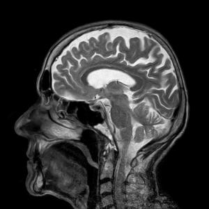
The main tool scientists use to look at brain structures is called magnetic resonance imaging or MRI, first introduced in Lesson 1. This technique allows scientists to take detailed pictures of the brain.
Unlike computerized axial tomography (CAT) scans that use x-rays, MRI is a non-invasive technique. MRIs use strong magnetic fields and radio waves to generate images of the brain like the one pictured on the left side of the screen.
When studying the development of the brain, researchers often ask the same child to come back for multiple scans as they grow. This allows researchers to see how the brain’s structure develops over time.
A special kind of MRI, called diffusion tensor imaging, or DTI, allows researchers to look specifically at white matter tracts in the brain.
The result of a DTI experiment is pictured on the right. Remember that white matter tracts are bundles of myelinated axons that act as information super highways in the brain.
In studying development, this technique can be useful to track which parts of the brain are myelinating and when. Then, for example, these results can be compared to behavioral tests to see if children’s developing abilities correlate with the refinement of white matter tracks in their brains.
B – Measuring Brain Activity—fMRI
A technique that is closely related to MRI, called functional MRI, or fMRI, is used to precisely pinpoint activity in the brain.
The technique fMRI works by measuring changes in the blood’s oxygen level, which varies in response to which area of the brain is active. Active regions of the brain require more oxygen, so by measuring oxygen levels in the blood, scientists can pinpoint where activity is happening in the brain.
This technique gives a more precise location of brain activity and is particularly useful for studying the activity of regions that are deep within the brain, such as structures involved in reward pathways.
Because fMRI does not measure brain activity directly and instead measures a biological response to brain activity, the signal is delayed. This means that it is difficult to accurately measure the timing of brain responses. So while fMRI is good for figuring where brain activity is happening, it’s not great for figuring out exactly when it is happening.
Brain activity changes rapidly, which can make it difficult for scientists to pinpoint exactly when different regions of the brain are active in response to which stimuli. For example, if scientists see brain activity in a specific area of the visual cortex when a person looks at a picture, it is hard to pinpoint if the brain activity occurred in response to the picture itself or something before or after the person saw the picture.
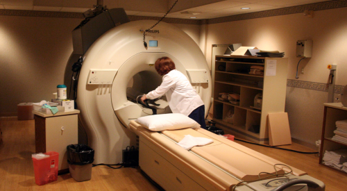
The physical constraints of the fMRI machine can also pose some challenges to researchers. For example, because the machines are so loud, it can be difficult to study the brain’s response to sounds. Children and adults also must stay perfectly still for a session, which can last between 15 minutes and an hour, depending on the study.
C – Measuring Brain Activity—EEG
Another technique that scientists use to measure brain activity is called electroencephalography, or EEG.
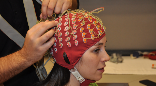
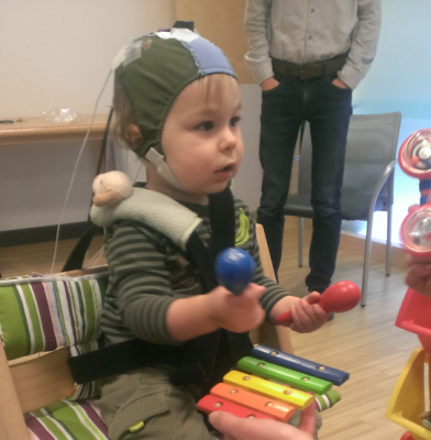
Unlike fMRI, which indirectly measures brain activity, EEG measures the electrical activity produced when many neurons in the brain fire together in sync. Finely tuned sensors stitched into a cap measure neural activity. Researchers place this cap over an adult or child’s head.
Because this technique directly measures brain activity, it allows researchers to pinpoint exactly when regions of the brain respond. For example, with this technique, researchers can get a glimpse of what is happening in the brain when a child responds to a touch or hears a familiar voice.
While EEG is silent and does not require the child to sit completely still, it cannot precisely locate where activity is happening in the brain. It just provides information about a general region.
D – Measuring Brain Activity—MEG
A third technique that scientists use to image the brain is called magnetoencephalography or MEG.
Location and Intensity
With this machine, scientists can directly measure both the location and intensity of brain activity. Like EEG, MEG is silent, non-invasive and harmless. MEG is a useful tool for determining what parts of the brain are active as people do certain tasks, like reading or listening to foreign language sounds.
MEG works by detecting the weak magnetic field produced by the chatter of neurons firing in unison within the brain. This type of information helps scientists understand how different parts of the brain work together.
Similar to EEG, the MEG machine, pictured on the left, has hundreds of extremely sensitive sensors located within the dome above the seat.
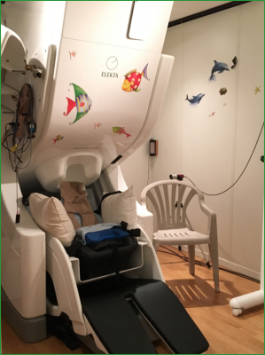
Infants are free to move their head in the MEG machine. Their head motions are tracked with a special cap, pictured on the right, that allows scientists to align their brain activity with a map of brain structure after the imaging session is over.
Later in the course, you will learn about studies that use all of these techniques to help scientists better understand how our brains develop as we finely tune them through everyday experiences.

Reflection Point
Why is it useful to study the brain?
If you could ask anything about the brain, what would you want to know?
 References
Berk, L. (2013). Child Development (9th ed.). Pearson.
Gigandet, X., Hagmann, P., Kurant, M., Cammoun, L., Meuli, R., & Thiran, J.-P. (2008, December 23). White matter connections obtained with MRI tractography [Online image]. In Estimating the confidence level of white matter connections obtained with MRI tractography. PLoS ONE 3(12). [Journal article]
Simon Fraser University – University Communications. (2014, April 15). Brain study. [Online image]
The B’s. (2009, April 17). MRI suite. [Online image]
EarlyEdU Alliance (Publisher). (2018). 2-3 Tools we use to study the brain. In Child Development: Brain Building Course Book. University of Washington. [UW Pressbooks]
