
1-3 Introduction to the Brain
A – Brain Development
Children’s brains grow faster in the first few months and years of life than they will at any other time.
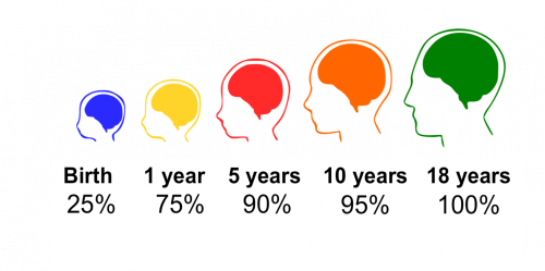
At birth, infants’ brains are about one-quarter the volume of adult brains. The rest of a newborn’s tiny body is not even close to one-quarter of their adult size. If it were, the average newborn in North America would weigh about 40 pounds.
Children’s brains continue to grow, and quickly. A recent study calculated the rate, or how fast, infant brains grow. By the end of their first year of life, children’s brains are already 75 percent of adult size. By 5 years of age, they grow to about 90 percent of adult size.
An important thing to note is that while 5-year-olds’ brains may be 90 percent of the size of adult brains, this does not mean that children’s brains are 90 percent finished by age 5. A 5-year-old has much, much more to learn.
For example, when 4 to 5 years old, children can only sometimes control their impulses and still need support from adults. The parts of the brain that control impulses, like the prefrontal cortex, still need more time and experience to develop. Scientists estimate that the brain doesn’t finish developing until well into the third decade of life. But by 5 years of age, children have most of the raw materials, like brain cells or neurons that build the brain.
Children’s brains are uniquely primed to learn from everyday experiences. At this stage, the brain is like a rough draft, ready for the experiences of life to continue shaping it into the specialized brain of an adult.
Session 2 will focus more on brain development in the first five years of life. But next will be a closer look at the brain and how it works.
Control and Communications Team
The brain is part of the nervous system. The nervous system is a bit like the body’s control center and communications team.
The nervous system controls both your voluntary functions like thoughts and actions, as well as your body’s involuntary functions like breathing and your heart beating.
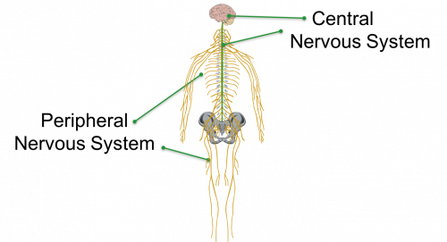
The nervous system has two main parts:
- The central nervous system, which is made up of the brain and the spinal cord.
- The peripheral nervous system, which includes all of the nerves that carry messages to and from the brain to the rest of the body.
The Nervous System and Neurons
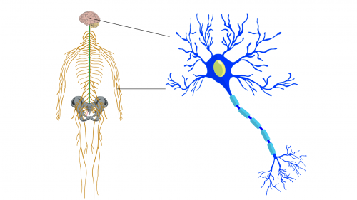
The nervous system is made up of neurons. Neurons are cells that have special features that allow them to quickly send messages throughout the body.
There are neurons in the brain—and a lot of them. The adult brain has about 86 billion neurons.
The neurons in our brains process information coming in from the body through the peripheral nervous system. From this information, neurons form our thoughts and direct our actions.
For example, if you see your friend in the grocery store, your brain processes the visual information. Several regions of the brain coordinate to recognize who this person is and to determine how you should react. The brain then directs the muscles in your arms and hand to wave and the muscles in your face to pull into a smile.
We also have neurons throughout the rest of our bodies. Some of these neurons sense the surrounding world—the sounds, smells, tastes, textures, touches, and temperatures—and translate these inputs into neural messages that travel to the brain.
Other neurons in the peripheral nervous system direct our muscles to contract, allowing our bodies to move and our heart to beat.
 Interactive: Parts of a Neuron
In this interactive, we look at some of the special features that enable neurons to send messages throughout the brain and body.
A Network of Neurons
At this point, it is important to note that neurons never work alone; each neuron is part of a network of other neurons.
Neurons transfer signals to other neurons through structures called synapses. A signal neuron may make thousands of synapses with other neurons.
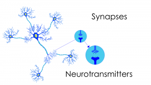
Once the signal reaches the end of the axon, it triggers the release of chemical messengers at the synapse. These chemical messengers are called neurotransmitters.
Neurotransmitters travel through the tiny space between neurons and dock in special receptors on the receiving neuron’s synapse. This initiates a weak electrical signal in the dendrites of the receiving neuron and the process begins again.
When someone is talking about making connections in the brain, they are talking about synapses. When we learn new information, the number or structure of synapses changes in our brains as a result of repeated experiences.
Myelin
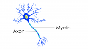
Many neurons have another feature that helps them to communicate information quickly. The name of this feature is myelin. Myelin is a fatty, insulating layer that wraps around axons. It helps electrical impulses to travel faster down the axon.
Here is an example: Have you ever grabbed a hot pan handle and then quickly let go a few seconds before you felt pain in your hand?
It turns out that the neurons that transmit the sensation of hot temperature to your brain have myelin on their axons, or are said to be myelinated. But the neurons that sense burning pain don’t have myelin. So the pain sensation arrives at the brain just a bit after you let go of the hot pan handle.
The brain has many myelinated axons. This allows neurons to rapidly and efficiently communicate.
B – More Brain Anatomy
Gray and White Matter
This image shows the brain as a whole. The outer part of the brain is called the cortex. The cortex is densely packed with the cell bodies of neurons.
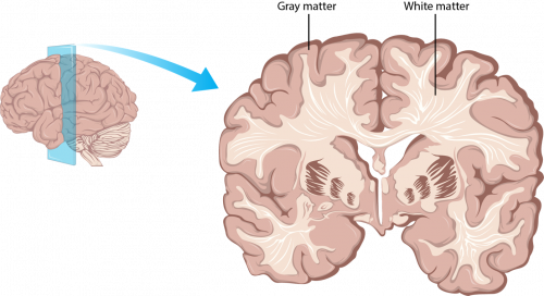
The cortex is actually slightly pink in color, but this part of the brain is often called gray matter. It is called gray matter because this dense tissue appears gray in magnetic resonance imaging (MRI) brain scans. Session 2 will focus more on brain imaging.
Beneath the gray matter are bundles of myelinated axons. This is called white matter. The fatty myelin coating on these bundles of axons appears white in MRI scans.
Many axons bundle together to form what are called fiber tracts. These fiber tracts are like superhighways for information, connecting different regions of the brain together. These fiber tracts allow neural messages to travel quickly from region to region.
Brain Networks
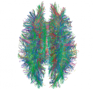
Using a special type of magnetic resonance imaging (MRI) called diffusion tensor imaging, or DTI, scientists can examine where different fiber tracts are located in the brain.
This image shows a three-dimensional model of white matter fiber tracts in the brain. We are looking straight down at the top of the head.
When looking at this image, can you imagine what the brain looks like, even though we are only looking at the white matter fiber tracts?
These information superhighways carry neural impulses rapidly throughout the brain. Everyone uses all of their brain constantly, not just a certain part, or a certain percentage.
Regions of the Brain
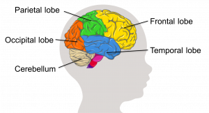
The brain has different functional and structural regions. These regions are called lobes. Each lobe is specialized for a particular set of tasks.
For example, the occipital lobe processes visual information. The frontal lobe is involved in reasoning and planning, the ability to control emotions, and motor control.
We use all of our brain constantly. Your brain works continuously, rapidly sending information between these different regions via fiber tracts and determining what you should say, do, and feel at any given moment.
Earlier, you imagined what the outer part of the brain looks like, just from seeing the underlying fiber tracts. Now, do the opposite: Looking at this illustration of the cortex, imagine what all of those fiber tracts that connect these different regions might look like.
C – The Working Brain
Brain Development and Learning
Our brains have a lot of different tasks to do. They take in information from our senses and process all of it simultaneously to make sense of the world. Based on all that input, our brains have to then coordinate how our bodies should respond.
This section covered some of the main regions in the brain and what their functions are. It also presented how neurons work to carry messages throughout our brains and bodies.
Throughout our lives, but especially as children, we train our brains to respond in different ways to different situations through the experiences that we have.
During the first few years of life, children’s brains are developing, growing, and learning at an incredible rate.
Session 2 will talk more about brain development in the first five years of life.
Video: How the Brain Works (3:54)
This animation is called How the Brain Works and reviews the information you’ve learned about the brain.
 References
Berk, L. (2013). Child development (9th ed.). Boston, MA: Pearson.
Cultivate Learning (Producer). (2017). How the brain works. University of Washington. [Video File]
Gigandet, X. (2008, December 23). White matter connections obtained with MRI tractography [Online image]. In Estimating the Confidence Level of White Matter Connections Obtained with MRI Tractography, PLoS ONE, 3(12), 1-9. [Journal article]
Mancall, E. L., & Brock, D. G. (2011). Gray’s clinical neuroanatomy: The anatomic basis for clinical neuroscience. Elsevier/Saunders
Thomason, M. E., & Thompson, P. M. (2011). Diffusion imaging, white matter, and psychopathology. Annual Review of Clinical Psychology, 7, 63-85.
EarlyEdU Alliance (Publisher). (2018). 1-3 Introduction to the Brain. In Child Development: Brain Building Course Book. University of Washington. [UW Pressbooks]
