
2-2 Stages of Brain Development
A – Stages of Brain Development
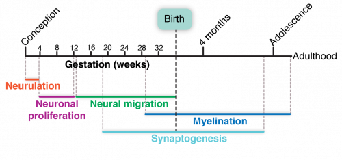
Brain development occurs over many stages. It begins soon after conception and continues to adulthood.
Let’s briefly highlight each step in brain development for a broad overview of the process.
Early Formation of Nervous System
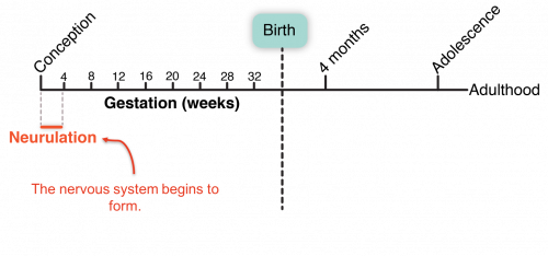
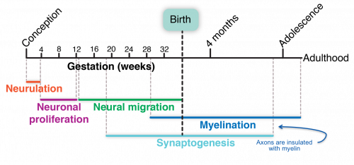
Brain development begins long before birth. In fact, the first stage of brain development, the formation of the nervous system, begins within days of conception.
As discussed in the last lesson, the nervous system is the body’s communication’s team. Networks of neural connections form an information superhighway. Rapid communication between the body and brain drives every single motion, intention, and thought. It is this network that allows people to learn and adapt to their ever-changing surroundings.
This early formation of the nervous system is called neurulation.
Knowing the term neurulation isn’t that important, but what is important to know is that the nervous system begins to form before most women know that they are pregnant. The developing nervous system is very susceptible to recreational drugs. Recreational drugs are psychoactive, meaning they are used to alter one’s mental state in a way that modifies emotions, perceptions, or feelings.
Psychoactive drugs change the way the nervous system communicates. When the nervous system is developing, these changes can cause the brain and the connections forming within the brain to develop in atypical ways. It is very important that women who are pregnant avoid recreational drugs and alcohol so that their baby’s brains develop without the interference of psychoactive substances.
In fact, the Centers for Disease Control and Prevention, known as the CDC, recommends that even women who are trying to become pregnant, or could become pregnant, avoid drinking alcohol. This is because most women don’t know they are pregnant for several weeks, and by that point the nervous system has already begun to form. The most recent estimate from the CDC is that up to one in 25 babies may have fetal alcohol spectrum disorder, which can lead to behavioral and intellectual disabilities.
From the earliest points in development, the brain is affected by its environment.
The Birth of Neurons
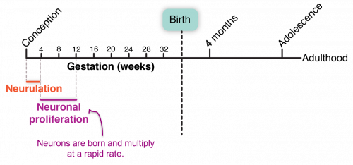
The next phase in brain development is the birth and rapid multiplication of neurons.
At birth, infants have many of the neurons that they will have as adults. Adult brains have about 86 billion neurons, so the developing brain has a lot of work to do as it generates billions of these specialized communication units.
Reviewing Neurons
Let’s briefly review what neurons are and how they work.
In the last lesson, we learned that the network of neurons in our bodies act as our personal communications teams. The neurons in the brain process information coming in from the body through the peripheral nervous system. From this information, neurons form thoughts and direct actions.
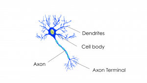
Neurons receive signals from other neurons through a network of intake fibers called dendrites. These signals travel down the dendrites, in the form of weak electrical currents, until they reach the cell body.
Once they reach the cell body, these signals, if they are strong enough, trigger the neuron to send or fire an electrical impulse.
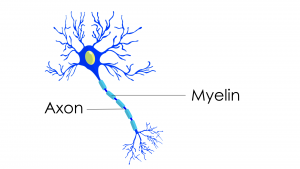
This electrical impulse travels down the length of the neuron’s axon, or output fiber.
Neurons transfer signals to other neurons through structures called synapses. A signal neuron may make thousands of synapses with other neurons.
This lesson will present more information on synapses later.
Neural Migration
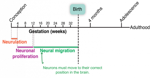
Most neurons are born deep in the central part of the developing brain.
They then have to move, or migrate, to their final position. This process is driven by tides of chemical signals that help newborn neurons find the right place in the brain. Getting to the right place is no easy task.
The brain is a complicated structure with many different functional zones. Next, we will take a look at some of those regions.
B – Rapid Brain Growth
Rapid Brain Growth
An infant’s brain grows at an incredible rate of about 1 percent a day, then slows slightly to 0.4 percent a day by the end of the first 3 months.
So far, we have mostly discussed brain development before birth. By the time a child is born, an infant’s brain has all of the different regions that it will have as an adult and most of the neurons. But the brain still has a huge period of growth and development to go through.
A child’s brain grows faster in the first few months and years of life than it will at any other time.
A recent study calculated the rate, or how fast, that infant brains grow. Researchers found that an infant’s brain grows at an incredible rate of about 1 percent a day, then slows slightly to 0.4 percent a day by the end of the first 3 months.
During this period of rapid brain growth, a lot is happening in the brain. One very important part of brain development is the formation of communication points, or synapses between neurons.
Video: Colors on the Piano (0:58)
This next video is Colors on the Piano. The video is approximately 1 minute long.
As you watch the following video, think about which brain areas the child may be using.

Video Debrief
Which part of the child’s brain regions do you think were active during the interaction on the video? (click to toggle expand or collapse)
Here are some of the regions of the brain we thought were active:
- Frontal lobe: planning what to do next, motor control
- Parietal lobe: sense of touch
- Occipital: vision
- Temporal lobe: learning language sounds
- Cerebellum: coordinating motor movements
- Limbic regions: remembering what to do with a piano
Process of Making Connections
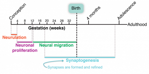
Neurons rely on synapses, or connection points between neurons, to communicate with each other.
The process of forming these connections, called synaptogenesis, extends from gestation to adolescence and even adulthood.
Next, we’ll learn more about what synapses are, how they form, and how they can change over time.
Synapses
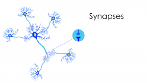
Previously, we talked about how neurons send messages down their axon, or output fiber, via weak electrical currents. What happens when that electrical current reaches the end of one neuron? How does one neuron share this message with the next neuron?
Messages move from one neuron to another across synapses. Synapses are similar to the point in a relay race where one runner passes the baton to the next runner. The runners themselves don’t touch, but they come very close, transferring the message, or the baton, from one runner to the next.
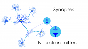
The weak electrical currents can’t cross through the tiny space between neurons. The message has to be translated into a different form. When the electrical current reaches a synapse at the end of the axon, it triggers the release of a burst of chemical messengers, or neurotransmitters. Neurotransmitters cross the tiny space between neurons, allowing the neural message to transfer from one neuron to the next.
Imagine the relay runners again. What if a runner threw a handful of confetti to the next runner instead of passing the baton? This is a little bit like what happens when the electrical impulse reaches the end of the axon. It triggers structures in the neurons to release a batch of chemical messengers.
These tiny chemical messengers sail through the space between neurons and dock in tiny receptor structures located on the receiving neuron. The receptor structures then translates this chemical message back into an electrical signal. And the electrical signal begins its journey down the dendrite toward the cell body.
Neurotransmitters
The brain has different types of neurotransmitters. This allows neurons to send specialized messages.
Most neurons release only one kind of neurotransmitter. In fact, many neurons are classified by the type of neurotransmitter they release. The two most common types of neurotransmitters transmit a signal that either increases the likelihood that the neuron will send a message of its own or decreases the likelihood.
Other neurons that release specialized neurotransmitters are only in certain brain regions. Some widely known specialized neurotransmitters are serotonin, dopamine, and epinephrine (also called adrenaline). While not the most common neurotransmitters in the brain, they do play a powerful role in modifying moods and regulating alertness.
Most drugs that people use to modulate moods change the effectiveness of neurotransmitters in the brain. This is the same for prescription drugs, such as anti-depressants, and recreational drugs, both legal, such as alcohol, and illegal, such as cocaine. All these drugs modulate neurotransmitter activity.
A child’s developing brain is particularly sensitive to changes in neurotransmitter dosage. Drugs that increase or decrease the dose of certain neurotransmitters can actually change how networks of neurons are wired together and how they function. This is why warnings exist for women who are pregnant about taking drugs, especially alcohol, that modify certain neurotransmitters.
Trillions of Connections
Neurons have different jobs in the brain. Some connect to just a few other neurons, while others connect to thousands of other neurons. Typically, one neuron will make many connections to another neuron. Regardless of whether neurons connect to one or many neurons, the average neuron makes 7,000 synaptic connections.
We are born with many of the neurons we will ever have—about 86 billion. Imagine that each of those 86 billion neurons then has to make 7,000 connections with other neurons. That is an incredible amount of synaptic development that has to occur.
It is estimated that in the cortex alone there are about 20 billion neurons and that those 20 billion neurons make about a trillion synapses per cubic centimeter of cortex.
Although we are born with most of our neurons, our brains have not yet made all of those trillions and trillions of connections. This allows us to continuously learn new things and become experts at living our own lives.
C – How Connections Develop
Synaptic development is sort of like a forest of young trees or saplings. All the trees are there, but they are small, and they haven’t filled in all their branches.
Neurons grow in a similar way. As the trees, or neural connections, grow, they mature into a dense network of connections—the ecosystem of the brain.
And this is a good thing because the connections forming in our brains are the result of what we learn and the experiences that we have. When you learn something new, you are shaping the way neurons in the brain share information.
Experiences Shape Connections
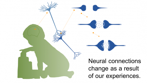
To a certain extent, which connections form between neurons, and how strong those connections are, is based on our experiences. The developing brain is establishing networks of neurons that work together.
The more often you have an experience, the more the neurons involved in processing that experience begin to work in unison. Perhaps you have heard the phrase: Cells that fire together, wire together. The idea behind this quotation is that the more often a certain neural pathway is stimulated, the stronger that neural pathway becomes.
Connections and communications between neurons grow stronger by increasing the number of synapses or increasing the size of existing synapses.
When we learn something new, and we practice what we have learned, we are shaping how the neurons in our brain connect and communicate.
With so much to learn in the first few years of life, our brains are forming new connections at an incredible rate. Your work as an early childhood educator is quite literally shaping the architecture of brain.
Experience Expectant
Scientists William Greenough and James Black described the way the brain makes connections by lumping them into two main categories.
The first category they called experience-expectant synaptogenesis. This describes the process of forming connections in the brain based on the experiences that every typically developing child will be exposed to. For example, the visual parts of the brain expect or require exposure to light and pattern to form the correct connections in the brain. The brain is pre-programmed to collect this information and form the appropriate connections in the brain.
While exposure to these types of environmental input or experience is required for typical brain development, barring an extreme circumstance, every child will form these connections without intervention or need for extra stimulation.
Experience Dependent
Greenough and Black referred to the second category of connection building as experience-dependent synaptogenesis.
This process describes the connections that form in our brains as a result of the unique experiences that we have in our lives. For example, we all learn to speak different languages.
The more exposure that we have to a particular experience or set of experiences, the stronger the connections between neurons become.
This is true of both positive and negative experiences in our lives. And this process starts from birth. Our experiences as children shape the way our brains are connected and set the stage for the rest of our lives.
Neuroplasticity
The ability to change the way neurons in the brain connect and communicate is called neuroplasticity. Just as you can mold plastic, you can shape the way that neurons in your brain network.
We can’t change all the connections in our brains. Some are fixed, but many others can be altered, at least to some degree, by the result of experiences. Scientists didn’t always know that the brain could be shaped by experiences.
It wasn’t until the 1960s that groundbreaking work by Dr. Marian Diamond and her colleagues, David Krech and Mark Rosenzweig, revealed that experiences can change the physical structure of the brain.
In their original experiments, Dr. Diamond and her colleagues looked at the brains of rats that either lived in cages that had lots of fun toys, like ladders, wheels, and balls, to keep them busy or lived in empty cages with nothing to do other than eat and drink.
The researchers found that the brains of rats that lived in cages with lots of opportunities to explore and be active were actually larger than those of the rats in the empty cages.
Dr. Diamond went on to discover that changes in experiences could lead to changes in the size of neurons and their number of connections.
Changes to Adult Brains
While the original studies on neuroplasticity involved rats, more recent studies have looked at how experiences can change the brains of humans as well, even adults.
One famous example of this is a study looking at the brains of taxi drivers in London, before widespread use of Global Positioning Systems (GPS) or smartphones.
Researchers used a brain imaging technique called magnetic resonance imaging to look at the size and shape of a region of the brain that is important for memory. Do you remember the name? This region is the hippocampus.
When the researchers looked at the hippocampus of taxi drivers, they found that a region of the hippocampus that is important for spatial memory was larger in the taxi drivers than in the brains of people who were not taxi drivers.
This indicates that even the adult brain has the capacity to make small changes to brain structures based on experiences and the demands of our everyday environments.
Video: My Love Affair with the Brain (6:07)
Listen for the information that Dr. Marian Diamond shares about the brain while you watch the next video.
To provide some information about the people making these world-changing discoveries, the scientists, we’ll watch this video trailer to a documentary film about the life and work of Dr. Marian Diamond. Dr. Diamond was a pioneer in the field of neuroplasticity. She died in the summer of 2017.
As you watch the video, consider:
- What did you hear Dr. Diamond say about the brain?
- How does this information apply to your interactions with children?
- How do you support children’s brain development?
Watch My Love Affair with the Brain from Bullfrog Films on YouTube.
Video Debrief
What are the implications for brain development? (click to toggle expand or collapse)
A couple points that Dr. Diamond made about the brain are:
- Diet, exercise, challenge, newness, and love can improve the brain.
- The brain is only 3 pounds, approximately.
The information in the video applies to everyday interactions with children because children are building neural connections and setting the foundation for years of learning.
You can support children’s brain development with any activity that involves responsive, social, back-and-forth interactions, or guided exploration of new experiences.
D – Brain Connections Activity
In this activity, you will take a closer look at the process of forming connections between neurons.
Images of brain neurons
The drawings in the interactive below show what neurons in the brain look like at different stages. These are drawings of what just a few neurons in the brain look like over the course of the first few years of life.
The dark triangular-shaped spots are neuronal cell bodies. The thinner lines are the axons and dendrites that carry messages between neurons. Within the first five years of life, the density of the connections between neurons increases rapidly.
 Interactive: Brain Connections by Age
In this activity, you’ll see a series of images that show what neurons in the brain look like at different stages. Inside of the interactive, arrange them in a way that you think best shows how the brain develops from the newborn brain to the brain of a six-year-old. Once you are done, click on the Check button to see how well you did. Note that you can attempt this activity multiple times.
Adapted by I-LABS from: LeRoy, CJ. The Postnatal Development of the Human Cerebral Cortex. Vol I- VIII Harvard University Press, Cambridge MA, Copyright 1939, 1975 by the President and Fellows of Harvard College.
 Activity Debrief
Were you surprised that a 6-year-old’s brain actually has fewer connections than a 4-year-old’s?
Why it matters
In the first five years of life, a child’s brain makes more connections than it will ever need; the brain overproduces connections. At age 5, a child’s brain has about two to three times the amount of connections that an adult’s brain has.
After over-producing connections, the brain begins to refine which connections to keep and which to discard. This process of refining the number of connections between neurons is called pruning. This pruning process is incredibly important for healthy brain development.
E – Pruning Connections
Pruning connections results in a thriving brain.
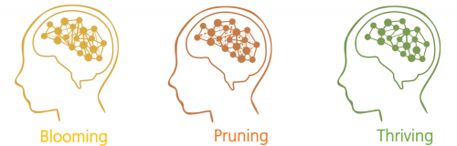
The brain prunes synaptic connections by reducing the number of connections it has, keeping the frequently used connections and eliminating the infrequently used ones.
This process is a little bit like the process of caring for a blackberry bush. At first there is a period of rapid growth, when the brain is blooming. During this time, the brain makes many extra connections. These extra connections actually make the brain less efficient.
Just like after a period of rapid growth in the spring, a blackberry bush can become gangly, with too many branches going in all different directions.
After the period of blooming in the brain, connections are refined, or pruned, based on the experiences that we have. Connections that we need and use are kept. Connections that we don’t need—ones that are actually making it more difficult for the brain to function—are removed.
The result of this process is a brain, or a blackberry bush, that is healthy and thriving. The branches, or connections, that are left are stronger and the brain is more efficient.
Throughout brain development, there are multiple periods of blooming and pruning. These bursts occur at different times and in different regions of the brain. Scientists think that these bursts of blooming and pruning align with sensitive periods in the brain. Sensitive periods are times when our brains are particularly open to new experiences and to learning.
Sensitive Periods for Language
The process of brain development is really the process of building the brain through experiences every day. Because the brain is developing so rapidly in the first few years of life, the experiences that people have as children are particularly influential.
The more often a child has an experience, positive or negative, the more likely that experience is to shape the connections forming in their brains.
Interactive: Critical Periods for Language
Use the image slider near the bottom of the interactive to explore this chart.

Reflection Point
Think about the kinds of experiences that children that you work with have on a daily basis. Consider these questions:
- What are children learning from those experiences?
- Would you change those experiences in any way to help support their learning?
F – Helping Neurons Communicate
Myelination
One final aspect of brain development to consider is the process of myelination. Myelination in the brain impacts how efficiently neurons communicate with each other.
Lesson 1 described myelination as the process by which neurons are coated in a fatty, insulating layer, called myelin, which wraps around axons. Myelin helps electrical impulses to travel faster down the axon.
Process Continues into Adulthood
Myelination is a long process that extends from before birth to early adulthood. This process bookends the period of rapid development in the brain. It starts before birth and extends well into people’s late 20s. Even as adults, biology and experiences continue to shape the physical structure of the brain.
When infants are born, few of their neurons are insulated with myelin. This means that their brains literally work slower than adult brains. Soon, the neurons begin to get their myelin coating, and the brain sends messages faster and faster.
The end result is that the brain works with much greater efficiency.
Significant Period for Myelination
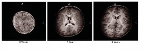
The most significant period of myelination occurs between mid-gestation and the second year of life. During this period, the density of myelin coating in the brain increases rapidly.
Sensory regions of the brain myelinate first, and then motor pathways, followed by regions of the brain that are important for higher cognitive functions.
Myelination reaches the prefrontal cortex between 7 and 11 months. However, the density of white matter continues to increase across development. Again, white matter is bundles of myelinated axons.
This is in part why infants require such a high fat diet—their bodies need that fat content to produce myelin and insulate neurons throughout their brains and bodies.
Myelination and Development
Myelination and white matter development correlates with the development of important skills. For example, one study found that the development of working memory and reading ability is coordinated with the maturation of white matter.
Scientists think that the development of skills across childhood may correlate with the time period when regions of the brain finish myelinating.
Other studies have indicated that, similar to neural connections, the density of white matter in different regions of the brain can also be modulated by experience. For example, pianists who practiced extensively as children have greater density of white matter in certain regions of the brain that are important for playing piano.
Biology + Experience Builds Brains
Over the course of childhood, we build our brains. This massive construction project is the result of both our biology and our experiences.
Our biology provides the neurons and the mechanisms to connect them, and it defines the structure of the brain. Our brains look very similar to the naked eye. Yet, at the microscopic level, we can see how our experiences influence how our brains are wired. Our experiences guide which neural connections form, become more efficient, and which will be removed. Our brains become adapted, or specially wired, for the tasks that we ask of them.
This combination of biology and experience contributes to all aspects of children’s development. During childhood, as children’s brains are growing and maturing at an incredible rate, they are eager to learn. As important adults in their lives, we can support children’s learning and brain development.
 References
Barkovich, A. J., Kjos, B. O., Jackson, D. E., & Norman, D. (1988). Normal maturation of the neonatal and infant brain: MR imaging at 1.5 T. Radiology, 166(1) 173–80.
Bengtsson, S., Nagy, Z., Skare, S., Forsman, L., Forssberg, H., & Ullén, F. (2005). Extensive piano practicing has regionally specific effects on white matter development. Nature Neuroscience, 8(9), 1148–1150.
Bennett, E. L., Diamond, M. C., Krech, D., & Rosenzweig, M. R. (1964). Chemical and anatomical plasticity of brain. Science, 146(3644), 610–619.
Berk, L. (2013). Child development (9th ed.). Pearson.
Bullfrog Films. (2017, April 4). My love affair with the brain. [Video]
Conel, J. L. (1939). The postnatal development of the human cerebral cortex (Vols. I–VIII). Harvard University Press.
Cultivate Learning (Producer). (2017). Colors on the piano. University of Washington. [Video File]
Diamond, M. C., Krech, D., & Rosenzweig, M. R. (1964). The effects of an enriched environment on the histology of the rat cerebral cortex. Journal of Comparative Neurology, 123(1),111–120.
Drachman, D. A. (2005). Do we have brain to spare? Neurology, 64(12), 2004–2005.
Gigandet, X., Hagmann, P., Kurant, M., Cammoun, L., Meuli, R., & Thiran, J.-P. (2008, December 23). Estimating the confidence level of white matter connections obtained with MRI tractography. PLoS ONE, 3(12). [Journal Article]
Gilmore, J. H., Lin, W., & Gerig, G. (2006). Fetal and neonatal brain development. American Journal of Psychiatry, 163(12), 2046.
Greenough, W. T., Black, J. E., & Wallace, C. S. (1987, June). Experience and brain development. Child Development, 58(3), 539–559.
Holland, D., Chang, L., Ernst, T. M., Curran, M., Buchthal, S. D., Alicata, D., … Dale A.M. (2014, October). Structural growth trajectories and rates of change in the first 3 months of infant brain development. JAMA Neurology, 71(10) 1266–74. [Journal Article]
Johnson, J. S., & Newport, E. L. (1989). Critical period effects in second language learning: The influence of maturational state on the acquisition of English as a second language. Cognitive Psychology, 21(1), 60–99.
Löwel, S., & Singer, W. (1992). Selection of intrinsic horizontal connections in the visual cortex by correlated neuronal activity. Science, 255(5041), 209–212.
Maguire, E. A., Gadian, D. G., Johnsrude, I. S., Good, C. D., Ashburner, J., Frackowiak, R. S. J., & Frith, C. D. (2000). Navigation-related structural change in the hippocampi of taxi drivers. PNAS, 97(8), 4398–403.
Nagy, Z., Westerberg, H., & Klingberg, T. (2004). Maturation of white matter is associated with the development of cognitive functions during childhood. Journal of Cognitive Neuroscience, 16(7), 1227-1233.
National Scientific Council on the Developing Child. (2008). The timing and quality of early experiences combine to shape brain architecture: Working paper #5. Harvard University. [PDF]
OpenStax College. (2013, May 23). 1511 The limbic lobe [Online image]. In Anatomy & Physiology. [eBook]
Seibel, N. L., Britt, D., Gillespie, L.G., & Parlakian, R. (2009). Preventing child abuse and neglect: Parent-provider partnerships in child care. Zero to Three Publishing.
Stoodley, C. J., Valera, E. M., & Schmahmann, J. D. (2012, January 16). Functional topography of the cerebellum for motor and cognitive tasks: An fMRI study. Neuroimage, 59(2), 1560–1570. [Journal Article]
Tau, G. Z., & Peterson, B. S. (2010). Normal development of brain circuits. Neuropsychopharmacology, 35(1), 147–168.
U.S. Department of Health and Human Services, Centers for Disease Control and Prevention. (2016). Alcohol and pregnancy. [Web Article]
Wagner, M. J., Kim, T. H., Savall, J., Schnitzer, M. J., & Luo, L. (2017, April 5). Cerebellar granule cells encode the expectation of reward. Nature, 544, 96–100. [Journal Article]
Yakovlev, P. I., & Lecours, A.-R. (1967). The myelogenetic cycles of regional maturation of the brain. In A. Minkowski (Ed.), Regional development of the brain in early life (pp. 3–65). Blackwell Scientific.
EarlyEdU Alliance (Publisher). (2018). 2-2 Stages of brain development. In Child Development: Brain Building Course Book. University of Washington. [UW Pressbooks]
