
2 Excitatory and Inhibitory Neurotransmitters
Synaptic potentials can be depolarizing (excitatory) or hyperpolarizing (inhibitory). The main ions involved in the generation of different synaptic potentials are Na+, K+ and Cl-. Their equilibrium potentials are approximately ECl ~ -90 mV, EK ~ -100 mV, and ENa ~ +70 mV. These values should be familiar! The rules for how changes in the permeability of the membrane to these ions affects the membrane potential are the same as those you learned in the membrane electricity session. Review those rules before proceeding if you are not comfortable with them! Recall that you can find most of them summarized in the separate chapter “Electric Keywords.”
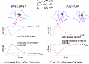
1. Excitatory postsynaptic potentials (EPSPs; Figure 3, left): Depolarizing excitatory postsynaptic potentials are generated by the opening of non-selective cation channels; current through these channels reverses at a potential of 0 mV, significantly more depolarized than the cell’s resting potential. Hence opening of these channels produces an inward current (referred to as an excitatory postsynaptic current or EPSC). This inward current produces the depolarizing excitatory synaptic potential, the EPSP.
2. Inhibitory postsynaptic potentials (IPSPs; Figure 3, right): Hyperpolarizing inhibitory postsynaptic potentials are generated by transmitter-gated ion channels permeable to either K+ or Cl-. Since the normal resting potential is more positive than the equilibrium potentials for K+ and Cl-, opening either of these types of channels will produce an outward current (an IPSC) generated by either outward K+ or inward Cl- flow, and this outward current will hyperpolarize the cell.
Excitatory and inhibitory synapses in action: The knee jerk reflex provides a nice example of the importance of EPSPs and IPSPs. You will learn about these spinal reflex pathways in more detail later; here we focus on the role of EPSPs and IPSPs. Extension of the lower leg involves increasing activity in the quadriceps muscles and decreasing activity in the hamstring muscles. The knee jerk reflex can be triggered when a reflex hammer imparts a small stretch to the quadriceps muscles; the stretch activates attached stretch receptors, producing action potentials in the sensory afferent nerve fibers innervating the stretch receptor. These afferent nerve fibers make excitatory synapses in the spinal cord both onto extensor motoneurons innervating the quadriceps and onto inhibitory interneurons in the spinal cord. Each of these afferent synapses produces EPSPs resulting in propagated action potentials. In the extensor motoneurons, APs lead to contraction of the quadriceps.
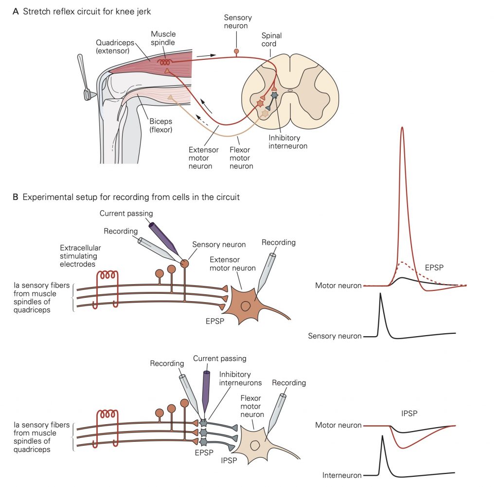
A. The stretch-receptor sensory neuron at the extensor (quadri- ceps) muscle makes an excitatory connection with an extensor motor neuron that innervates this same muscle group. It also makes an excitatory connection with an interneuron, which in turn makes an inhibitory connection with a flexor motor neuron that innervates the antagonist biceps femoris muscle group. Conversely, an afferent fiber from the biceps (not shown) excites an interneuron that makes an inhibitory synapse on the extensor motor neuron.
B. This idealized experimental setup shows the approaches to studying the inhibition and excitation of motor neurons in the pathway illustrated in part A. Above: Two alternatives for elicit- ing excitatory postsynaptic potentials (EPSPs) in the extensor motor neuron. A single presynaptic axon can be stimulated by inserting a current-passing electrode into the sensory neuron cell body. An action potential in the sensory neuron stimulated in this way triggers a small EPSP in the extensor motor neuron (black trace). Alternatively, the whole afferent nerve from the quadriceps can be stimulated electrically with extracellular electrodes. The excitation of many afferent neurons through the extracellular electrode generates a synaptic potential (dashed trace) large enough to initiate an action potential (red trace). Below: The setup for eliciting and measuring inhibitory poten- tials in the flexor motor neuron. Intracellular stimulation of a sin- gle inhibitory interneuron receiving input from the quadriceps pathway produces a small inhibitory (hyperpolarizing) postsyn- aptic potential (IPSP) in the flexor motor neuron (black trace). Extracellular stimulation recruits numerous inhibitory neurons and generates a larger postsynaptic IPSP (red trace). (Action potentials from the sensory neuron and interneuron appear smaller because they were recorded at lower amplification than the motor neuron action potentials).
EPSPs in the inhibitory interneurons increase the rate at which they generate action potentials. The inhibitory interneurons make inhibitory synapses with the flexor motoneurons innervating the hamstrings. Thus, action potentials in the inhibitory interneurons lead to IPSPs that will decrease activity in the flexor motoneurons (the motoneurons exhibit some spontaneous activity). The decreased flexor motoneuron activity decreases activity of the hamstrings.
Together, the increase in activity of the quadriceps and decrease in activity of the hamstrings leads to extension of the knee. The opposing changes in activity in the two muscle types in response to activation of the same stretch receptors illustrates the importance of having both excitatory and inhibitory synapses.