5 Chemical and Electrical Synapses
The second major form of synaptic transmission is mediated by electrical synapses. At an electrical synapse, signals are transmitted from one cell to another by direct current flow through structures called gap junctions (Figure 9). Electrical synapses are less common than chemical synapses in our brain, but are often found in sensory systems (like the retina) and in non-neural tissues like epithelia, the liver, and heart. They play an especially important role in synchronizing groups of cells. In nonneuronal systems, only the phrase gap junction rather than the word synapse is used for such common cell-to-cell communication. In evolutionary terms, gap junction structures are ancient forms of cellular communication.
Gap junctions provide a non-selective conductance path between the two cells, permitting exchange of ions, second messengers, and small molecules between the cells. They are often identified based on their ability to pass small tracer molecules such as the yellow dye illustrated in Figure 10; here the dye was injected into one of the cells, and passed, via gap junctions, into the others. Because gap junctions provide a non-selective path for current to flow, they tend to equalize the voltage in the two cells. This is illustrated in Figure 11. If V1 > V2, current will flow from cell 1 to cell 2 and bring the voltages closer together. Vice-versa if V2 > V1. This property makes electrical synapses particularly good at synchronizing the electrical activity in populations of cells, a property essential for cell-to-cell propagation of action potentials in cardiac muscle.
Indeed, many cardiac diseases are marked by a disruption of normal patterns of gap junctions between the cells. Unfortunately, we have few good (i.e. specific) drugs that act on gap junctions. This hampers study of their function and the treatment of disorders associated with them.
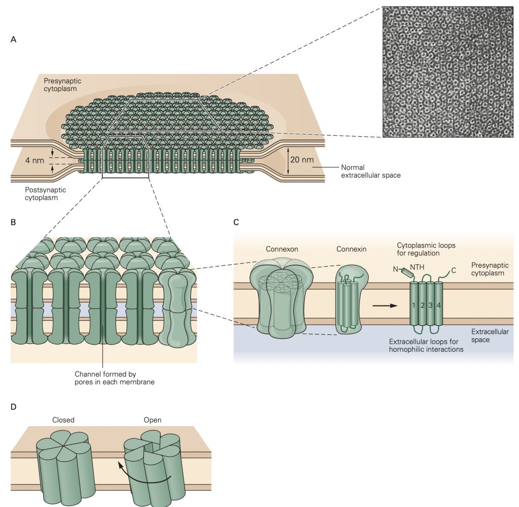
C. Each hemichannel, or connexon, is made up of six identical subunits called connexins. Each connexin is approximately 7.5 nm long and spans the cell membrane. A single connexin has intracellular N- and C-termini, including a short intra- cellular N-terminal α-helix (NTH), and four membrane-spanning α-helixes (1–4). There are regions of similarity in the amino acid sequences of gap-junction proteins from many different kinds of tissue. These include the transmembrane helixes and the extracellular regions, which are involved in the homophilic matching of apposite hemichannels. D. The connexins are arranged in such a way that a pore is formed in the center of the structure. The resulting connexon, with a pore diameter of approximately 1.5 to 2 nm, has a characteristic hexagonal outline, as shown in part A. In some gap-junction channels the pore is opened when the subunits rotate approximately 0.9 nm at the cytoplasmic base in a clock- wise direction.
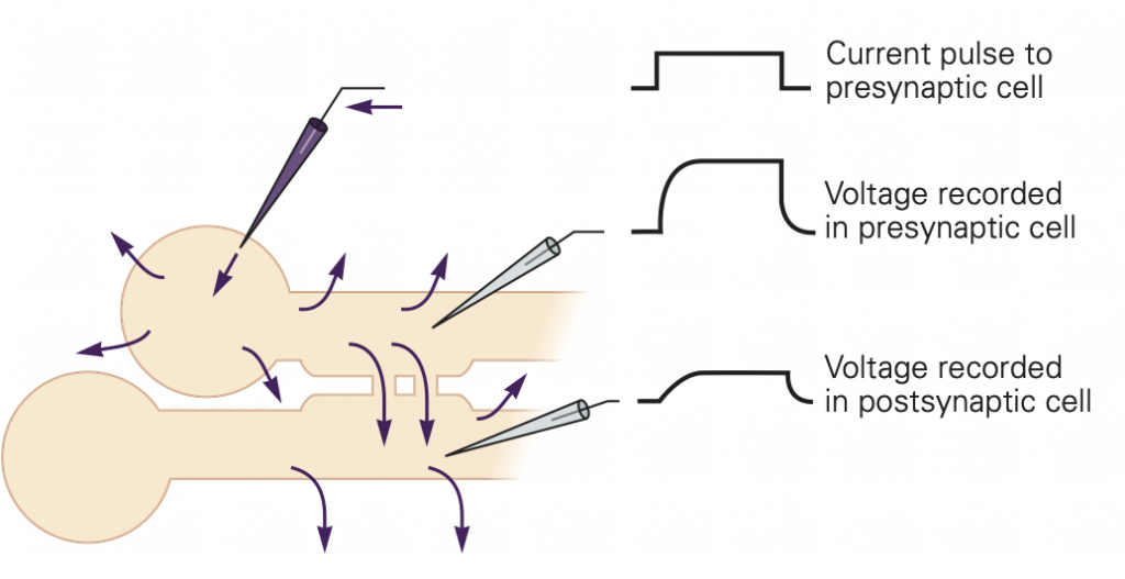
The electrical synapse relies on current from one cell causing a voltage change in the connected cell. For this form of synaptic transmission to be effective, the two cells must have similar resistances. If the cells have very different resistances, current passed from the high resistance cell to the low resistance cell will not be very effective in changing its voltage. As an analogy, think of pressing a hot needle up against a cannon ball; the needle doesn’t do much to heat the cannon ball because of its small size. A hot cannon ball, pressed up against the second cannon ball, would be much more effective. These reasons, for example, make gap junctions unsuitable at our neuromuscular junctions where motoneuron axons are small compared to the muscle cells that they innervate.