Nervous Tissue
SESSION OBJECTIVES
- Know the basic types of neural tissue: Central nervous system (CNS), Peripheral nervous system (PNS), Special senses.
- Know the two basic divisions of CNS: Gray matter: nerve cell bodies, dendrites and axons (neuropil), and glial cells. White matter: the axons of the neurons in the gray matter.
- Recognize the basic cell types in the nervous system: neurons, Glia and Support cells, Non-neural cells.
- Identify the basic parts of Neurons: axons, dendrites, soma, synapses, neuropil.
- Identify Myelin in PNS and CNS and know the cells that make this in PNS and CNS.
- Know the components of peripheral nerve and identify them in sections.
- Understand the process of Axoplasmic transport and how this impacts regeneration of axons.
OPTIONAL PRE-CLASS MATERIALS FOR THIS SESSION:
- Skim the section titles, bolded terms, and image captions from Junqueira’s Basic Histology, Chapter 9 (Click here for FMR library resources) to fill in any knowledge gaps you need.
NEURAL TISSUE TYPES (Session Objective #1): The nervous system has three basic types of neural tissue:
I. Central nervous system (CNS)comprised of the brain and spinal cord
-
-
-
- Most CNS cells are derived from the neural tube.
-
-
II. Peripheral nervous system (PNS) comprised of peripheral ganglia and nerves.
-
-
-
- Most PNS cells are derived from the neural crest.
-
-
III. Special senses (derived from both the Neural tube and/or the neural crest, and from specialized “placodes”) will be discussed in its own chapter.
In the next section the cells of the CNS will be described, and the following section describes the cells of the PNS.
I. CENTRAL NERVOUS SYSTEM (CNS)
DIVISIONS (Session Objective #2)
The CNS can be divided into gray and white matter
-
- Gray matter consists mostly of nerve cell bodies, dendrites and axons (together called neuropil), and glial cells (e.g. cerebral cortex, or central horns of the spinal cord).
- White matter is made up of the axons of the neurons in the gray matter (e.g. Corpus callosum of the cerebrum, and the tracts of the spinal cord.
CELL TYPES (Session Objective #3): There are three basic cell types in the nervous system:
1. Neurons
2. Glia and Support cells
3. Non-neural cells
CNS NEURONS (Session Objectives #3 and #4)
Neurons are electrically excitable cells that are highly polarized structurally and which have the capability of communicating signals (often at great distances) by means of action potentials propagated along axons, and transmitted to other neurons and cells at special junctions called synapses.
Knowledge check: Identify all of the structures of a CNS neuron:
- Cell body/soma:
- Neuron cell bodies contain a large, rounded nucleus with a prominent nucleolus.
- Neurons actively synthesize proteins to maintain their long processes and to produce neurotransmitters. Their cell bodies therefore contain large amounts of rough ER. The ribosomes stain strongly with the classic histological Nissl stain and are sometimes referred to as Nissl bodies or Nissl substance (these terms are somewhat outdated, but are still sometimes used in neuropathology, so you should be aware of them).
- Dendrites
- Dendrites are processes that receive stimuli from other neurons at synapses and conduct these to the cell body.
- Most neurons in the CNS have many dendrites and are therefore called multipolar. If a neuron only has one axon and one dendrite, it is called bipolar. There are only a few types of bipolar neurons in the CNS.
- Dendrites are usually highly branched, like a tree. (Dendrite = tree).
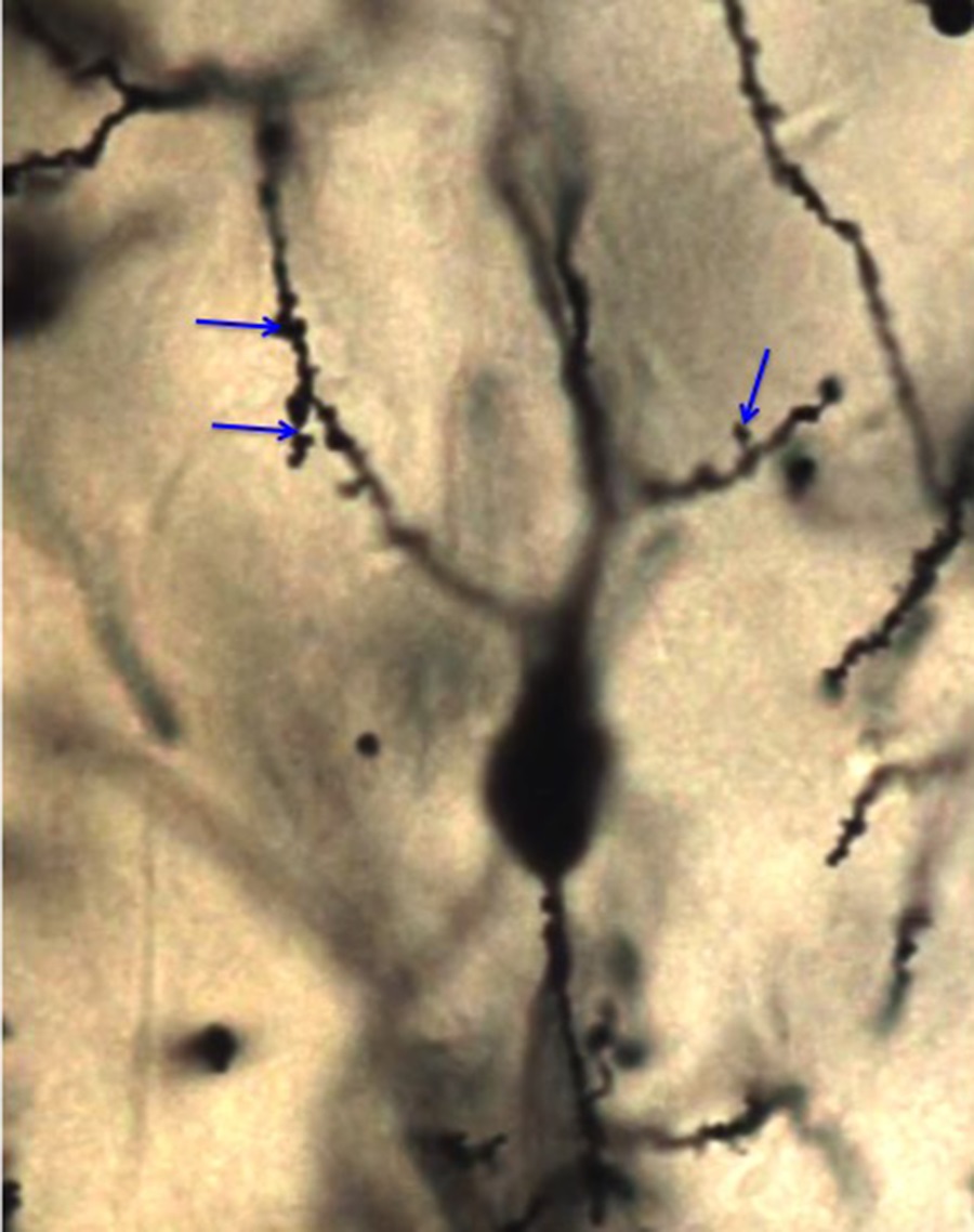
- In many neurons, dendrites are covered with many small projections on their surface, which are called spines. Spines are sites of synaptic contact, and are discussed more below in the Synapse section, but synapses can also be on the non-spiny parts of dendrites.
- Dendritic cytoskeleton contains microtubules and associated proteins (MAPs). Neuron-specific beta tubulin is used to identify neurons by immunolabeling techniques. MAP2 stabilizes the microtubules in dendrites.
- Dendrites have ribosomes and protein synthesis.
- Axons
- Most neurons possess a single axon, a process that conducts action potentials away from the cell body.
- The neuron’s axon arises from the cell body at the axon hillock, where action potentials are initiated.
- Axons are smooth processes (without spines) and usually do not branch near the cell body, but may give rise to collateral branches.
- Often, axons branch profusely at their end at terminal arbors.
- Axon cytoskeleton has prominent microtubules and intermediate filaments made of neurofilament protein. Neurofilament protein binds silver, and so classic histological silver nitrate stains for axons were commonly used to describe axon pathways.
- Axoplasmic transport (Session Objective #7): The axon of a neuron can be many centimeters and even meters in length. There are no ribosomes in axons and so no protein synthesis. To get cellular components to the axon from their point of synthesis in the cell body, neurons have a specialized transport system called axoplasmic transport. There are two basic components to axoplasmic transport:
- Fast transport-400 mm/day
- mostly vesicles
- vesicles move along microtubule tracks
- mediated by kinesin in the anterograde direction
- mediated by dynein in the retrograde direction
- microtubule disrupting drugs cause neuropathy
- Slow transport-1 mm/day.
- larger cell components, cytoskeleton and cytoplasm
- limits the growth rate of regenerating axons
- Fast transport-400 mm/day
- Synapses
- Synapses are sites of chemical communication usually found between the end of an axon and a cell body or dendrites of another neuron. Synapses are also found between dendrites (a dendro- dendritic synapse) and between axons (an axoaxonic synapse).
- In most synapses, chemical neurotransmitter molecules are released from the axon terminal (also called a bouton) and diffuse across a small synaptic cleft to active specific neurotransmitter receptors on the postsynaptic dendrite.
- Synapses are pretty small and so only recently has it been possible to visualize them with the light microscope; most of what we know about their structure comes from EM studies.
- Synapses are junctions between cells, and so they have a presynaptic side and a postsynaptic side; the two sides have specialized functions and are therefore structurally distinct.
- Presynaptic specializations:
- Synaptic vesicles contain the neurotransmitter molecules and fuse with the postsynaptic membrane to release them.
- active zone where the vesicles fuse to release transmitter
- The periactive zone is next to the active zone, and is the place where the membrane is endocytosed via clathrin coated vesicles for refilling with transmitter molecules; this process is called recycling.
- Postsynaptic specializations:
- Neurotransmitter receptors: (ligand gated or G-protein coupled receptors for the neurotransmitters released from the presynaptic axon terminal).
- The postsynaptic density (PSD) is a region of electron dense material in the postsynaptic cell immediately adjacent to the presynaptic vesicles. It is composed of proteins that concentrate the neurotransmitter receptors to this spot. There are also adhesive proteins in the PSD that attach the postsynaptic cell to the presynaptic cell.
- Presynaptic specializations:
- Spines:
- small protuberances from the surfaces of the dendrites of many neurons.
- usually receive excitatory input.
- their size and shape changes over time and correlate with synaptic strength
- new spines are made during learning and their retention correlates with the retention of specific memories. (Memories are made of these).
- Electrical synapses: Some synapses are actually gap junctions and transmit electrical signals without a chemical neurotransmitter. These do not have the typical pre- and post-synaptic specializations.
GLIAL CELLS AND OTHER SUPPORT CELLS DERIVED FROM THE NEURAL TUBE (Session Objective #3)
Glial cells, aka “Neuroglia” (= “nerve glue”) occupy the space between neurons, separating neurons from each other and from blood vessels. Three types of neuroglial cells have been classically identified in the CNS: astrocytes, oligodendrocytes and microglia.
- ASTROCYTES
- Astrocytes (astroglia) are star-shaped (“astro”) cells.
- Astrocytes are not all the same and neuroscientists are just beginning to figure out how much diversity there is in these cells; however, traditionally they have been divided into two basic types: Protoplasmic astrocytes (in the gray matter of the brain and spinal cord) and Fibrous astrocytes (in the white matter).
- Functions of Astrocytes:
a. transport materials between neurons and blood vessels,
b. give structural rigidity to the CNS,
c. regulate the composition of the extracellular space around neurons,
d. store energy (mainly as glycogen),
e. secrete growth factors
-
- Astrocyte cytoskeleton is primarily microtubules and intermediate filament proteins made up of “glial-fibrillary acidic protein” or GFAP; astrocytes increase GFAP expression with damage or when nearby neurons are under stress and so this can be an early marker of neural damage.
- The most deadly brain tumors resemble astrocytes and are thought to derive from astrocytes. These are called astrocytomas and in their more aggressive stage, glioblastoma multiforme.
- OLIGODENDROCYTES (Session Objective #3 and #5)
- Oligodendrocytes (oligodendroglia) are most abundant in the white matter of the CNS, where they form the myelin sheaths of axons.
- Oligodendrocytes make myelin, a compact sheath made of many layers of cell membrane wrapped around the shaft of axons.
- Oligodendrocytes wrap only a short stretch of each axon, but they can wrap myelin around up to 50 axons.
- The gaps between the myelin sheath made by each oligodendrocyte are called the nodes of Ranvier.
- Myelin increases the effective space constant of the axon membrane so that the action potential skips from node to node (saltatory propagation) and this results in a great increase in conduction velocity.
- EPENDYMAL CELLS
- Ependymal cells are an epithelium-like layer lining the cavities (ventricles) of the brain and the central canal of the spinal cord. These cavities contain the cerebrospinal fluid (CSF).
- The apical surfaces of these cells are covered with cilia, which help to circulate the CSF.
- In a few regions of the brain (the lateral wall of the cerebral ventricles and the hippocampus), there are specialized neural stem cells mixed in with the ependymal cells. These neural stem cells have a single primary cilium and generate new neurons throughout your life. In the hippocampus, these new neurons are important for new memories.
- CHOROID PLEXUS
- Choroid plexus is the term given to folds of ependyma (choroidal villi) extending into the ventricles of the brain.
- The plexus contains many blood vessels and is covered by cuboidal epithelium that secretes the CSF.
- The CSF acts as a cushion for the brain, suspending it in fluid within the dura.
- A blood-CSF barrier is established by the zonula occludens between the epithelial cells.
CELLS NOT DERIVED FROM THE NEURAL TUBE RESIDING IN THE CNS (Session Objective #3)
- MICROGLIA
- Microglia are small (hence “micro”), dense, highly branched cells with processes covered with thorn like projections.
- Microglia are migratory and phagocytic and are derived from circulating macrophages. You will hear more about macrophages later in the course.
BLOOD VESSELS and the BLOOD-BRAIN BARRIER
- The brain has a rich blood supply, but the capillaries in the brain have a restricted permeability that reduces exchange of large molecules between the blood and the brain cells, called the blood-brain barrier, or BBB.
- The BBB is due to tight junctions (aka zonula occludens) between capillary endothelial cells.
- The blood-brain barrier prevents most metabolites in blood from freely entering the brain. Essential metabolites in blood are transported into the brain across the blood-brain barrier.
- The BBB makes it difficult to get systemically administered antibiotics into the brain, and so brain infections can be hard to treat.
- Certain regions of the brain, like the hypothalamus, have a permeable BBB so that they can “sample” the blood for regulatory molecules.
MENINGES
- Meninges are the fibrous, connective tissue coverings of the CNS.
- There are three layers of meninges:
- Outer dura mater (“tough mother”),
- Middle arachnoid layer (“spider mother”),
- Inner pia mater (“soft mother”).
- The subarachnoid space between the arachnoid layer and pia mater is bridged by arachnoid trabeculae and filled with CSF.
- The pia mater is a thin layer of tissue that coats the outer surface of the CNS and fuses with the arachnoid trabeculae.
II. PERIPHERAL NERVOUS SYSTEM (PNS)
NEURONS AND GANGLIA
- The PNS includes all the neural tissue that is not located in the brain or spinal cord (or the special senses).
- Most neuronal cell bodies that lie outside the CNS are grouped into ganglia: a collection of neurons and associated satellite glia surrounded by a capsule of connective tissue.
- Sometimes the neurons are not collected in ganglia but are instead in more diffuse plexuses. In the gut, for example, you will learn about Meissner’s plexus and Auerbach’s plexus. These are more like nerve networks.
- The major types of ganglia in the PNS are:
-
- Dorsal root ganglia (DRG): Cranial (head) and spinal (rest of body) ganglia
-
-
- These contain the cell bodies of sensory neurons that carry somatosensory information from the periphery to the central nervous system.
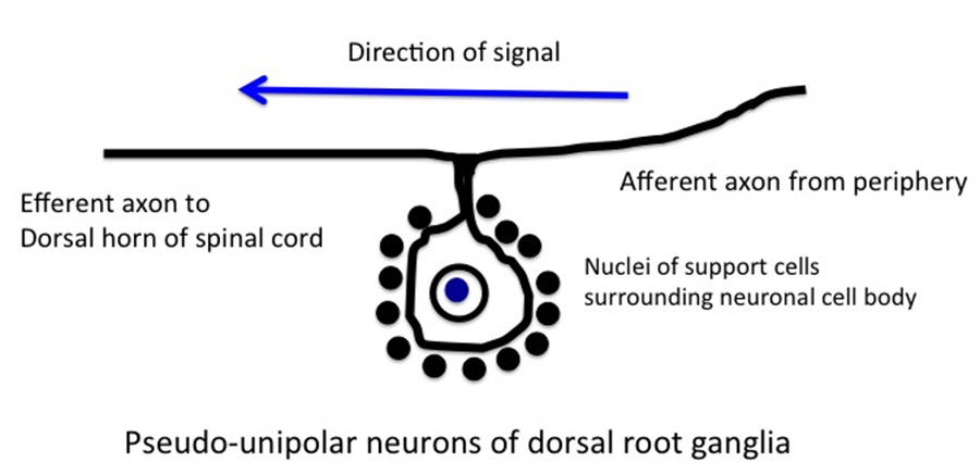
- The DRG neurons have a unique morphology; each has a central and a peripheral process connected to a single bifurcated process connecting the cell body. Since the neurons have only a single process emanating from the cell body, but then two processes after the bifurcation, they are sometimes called pseudounipolar.
- These neurons mediate sensations of touch, pressure, pain and position sense, via their connections with specialized sense organs in the skin (e.g. Pacinian corpuscles) and muscles (spindles). You will hear more about these receptor organs in the skin and muscle sessions.
- Two Types of Autonomic ganglia:
-
1. Sympathetic:
-
-
-
- The sympathetic ganglia are formed from enlargements of the spinal roots that contain the cell bodies of the postganglionic nerves of the sympathetic autonomic nervous system.
- Unlike the DRG neurons, the sympathetic ganglia neurons are multipolar neurons with dendrites and a single axon. Because of their dendrites, the neurons of sympathetic ganglia are less tightly packed than the DRG neurons; this difference makes it easy to distinguish these two types of ganglia from one another.
-
-
2. Parasympathetic:
-
-
-
- The parasympathetic neurons are collected in ganglia closer to the organs that they innervate, called intramural ganglia. These are small ganglia that contain a few thousand neurons (the ciliary ganglion for example contains only about 2500 neurons).
- Parasympathetic neurons come in a variety of shapes depending on the ganglion.
-
-
NOTE: Although many of the neurons of the PNS have their cell bodies outside the CNS in ganglia, the axons of the motor neurons that project to the muscles, or the preganglionic autonomic neurons, are also considered PNS, even though their cell bodies are inside the CNS (spinal cord). You will learn much more about these in your Neuro course.
Knowledge Check: Identify the Nuerons:
PNS GLIA
- SCHWANN CELLS (Session Objective #3 and #5)
- Schwann cells are the principal glial cells of the PNS. They are all derived from the neural crest. There are two types:
1. Myelinating Schwann cells wrap axons of the peripheral nerves in myelin much like the oligodendroglial cells of the CNS.
a. Like the oligos in the CNS, an individual Schwann cell covers only a short stretch of the axon of a neuron.
b. The gaps between the myelin from each Schwann cell are called nodes of Ranvier.
c. Unlike the oligos in the CNS, a Schwann cell covers only a single axon, while an oligo can coat many different axons.
d. The number of myelin wraps correlates with the size of the axon. A specific growth factor, called glial growth factor (or GGF) released from the axons, stimulates the myelin wrapping and the larger the diameter of the axon, the more GGF, and the greater number of myelin wraps.
e. Although CNS and PNS myelin look pretty much the same histologically, and are both composed largely of lipid, but there are some important molecular differences. The layers of adjacent membrane are bound by an interaction between proteolipid protein and myelin basic protein (MBP) in the CNS and by P0 protein and MBP in the PNS.
f. Diseases that cause the loss of myelin (demyelination) lead to changes in sensation and weakness due to conduction failure in the nerves. Two demyelinating diseases affect the CNS and PNS myelin differentially: Guillain–Barré is thought to be an autoimmune disease of PNS myelin and multiple sclerosis (MS) is a disease of CNS myelin. Differences in myelin composition (above) may explain the differential pathology.
2. Non-myelinating Schwann cells also envelope small axons in the peripheral nerves, though they do not make myelin.
a. The non-myelinating Schwann cells can surround several axons, instead of just one.
b. Non-myelinating Schwann cells also surround the axon terminals of the motor neurons and their synapses with muscles.
c. The zone of apposition between the adjacent Schwann cell membranes as it surrounds an axon is called the mesaxon.
NOTE: Your text calls Schwann cells “neurolemmocytes” or “lemmocytes.” Other Histo texts (e.g. Netter) use the term “neurilemmocytes.” These terms are no longer used very much in the scientific literature (2 citations in PubMed for neurolemmocytes), though are occasionally still used in pathology textbooks. It is not clear to me why Histo texts still use them. They will not be used in this course, but you might want to be aware of their existence.
- SATELLITE CELLS
- Satellite cells are the glia in ganglia
- They surround the neurons of the ganglia
- They are similar to Schwann cells, and some people group them all together
- Like Schwann cells, they are derived from the neural crest.
CONNECTIVE TISSUE SHEATHS AND PERIPHERAL NERVES (Session Objective #6)
The axons and Schwann cells of peripheral ganglia and nerves receive their structural support from connective tissue;
- Nerve fibers in peripheral nerves are grouped in bundles (fascicles).
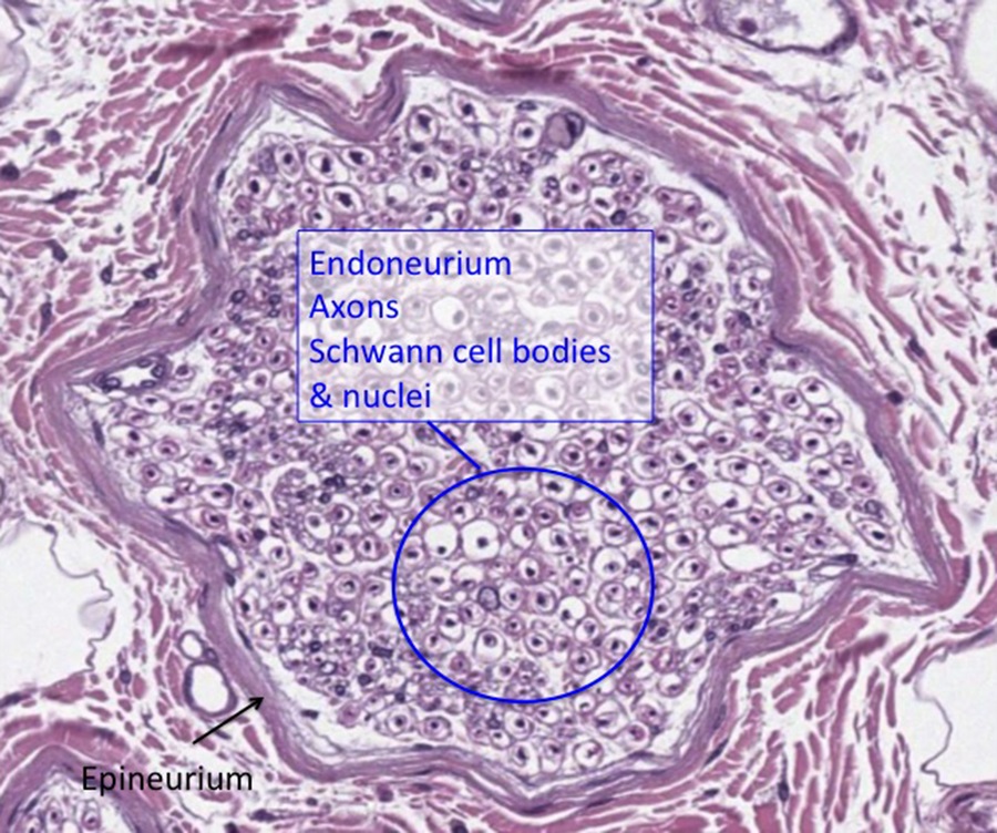
- The endoneurium surrounds individual Schwann cells and their enclosed axons.
- The perineurium surrounds bundles of nerve fibers in a nerve.
- The epineurium ensheaths groups of nerve fiber bundles, making the outer connective tissue sheath of the nerve.
Identifying peripheral nerves in cross-section and longitudinal section:
In the histology atlas and practice exercises below, you will work on identifying peripheral nerves.
Briefly, the image below shows a peripheral nerve in cross-section where you can see the epineurium, axons, endoneurium (surrounding each axon), and the nuclei of the Schwann cells:
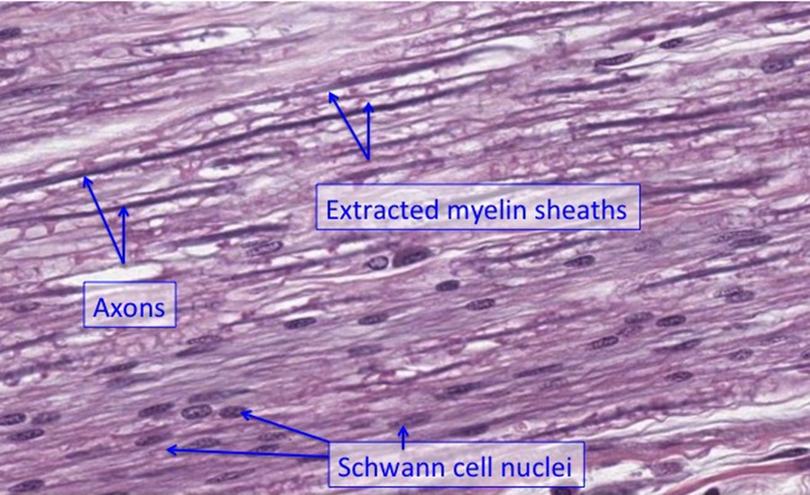
A peripheral nerve in longitudinal section may initially appear to be dense regular connective tissue of a tendon, but notice the increased cellularity (and rounder nuclei of the Schwann cells compared to fibroblasts) and the greater amount of white “spaces” that is myelin extracted from the section during processing. The long, fiber-like structures are basophilic axons of neurons rather than eosiinophilic collagen fibers as would be seen in and H&E stain of dense regular connective tissues (see below):
III. INJURY AND REGENERATION IN THE NERVOUS SYSTEM
- RESPONSE TO INJURY
- When the axon of a neuron is severed (axotomy) in a nerve injury, the part of the axon still attached to the cell body of the neuron can survive, but the part of the axon distal to the cut site degenerates by a process known as Wallerian degeneration.
- After axotomy, the neuron cell body undergoes changes in histological appearance called the “cell body response” or chromatolysis. The hallmarks of chromatolysis include:
- increase in cell size,
- an eccentric nucleus,
- and the loss in the Nissl staining due to dispersal of the stacked RER, hence the “lysis” in the “chromato.”
- These changes may be due to the accumulation of cytoskeletal proteins in the cell body that would normally be transported down the axon.
- In the CNS (e.g., spinal cord injury), the cut sites of the axons do not regenerate and so function is not restored distal to the lesion. However, some recovery is possible after spinal cord injury, because the cord is rarely fully transected.
- In the peripheral nervous system, axons in peripheral nerves can regenerate and reinnervate targets, particularly if the nerve is crushed rather than transected.
- REGENERATION
- After an axon in a peripheral nerve is cut, and the distal portion has undergone Wallerian degeneration, the proximal end sprouts a growth cone; this specialized structure has extensive fine processes that help guide the regenerating axon to the correct targets.
- In a crush injury to a nerve, the connective tissue sheaths are still intact, and the regenerating axons extend along lines of Schwann cells back to their original targets.
- When the regenerating axon reaches the target, for example a muscle cell, it forms a synapse at the original location, guided by the extracellular matrix.
- In a cut injury, the growing axons often cannot find the correct target and grow into a tangle of fibers called a neuroma.
HISTOLOGY ATLAS AND PRACTICE EXERCISES
Slide 1. Peripheral Nerve
The nerve shown here has been cut in cross section. It is stained with hematoxylin and eosin.
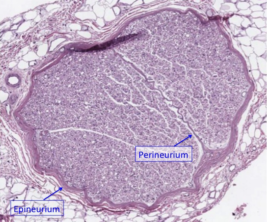
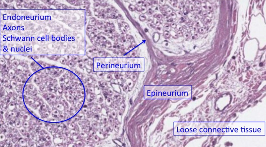
The nerve is covered with a dense connective tissue- the epineurium. Smaller sheets of connective tissue variably separate compartments of the nerve = perineurium. In this specimen above, the nerve is split into two main fascicles or bundles by the perineurium. Finally, there are delicate bits of connective tissue that separate individual axons and associated Schwann cells = endoneurium.
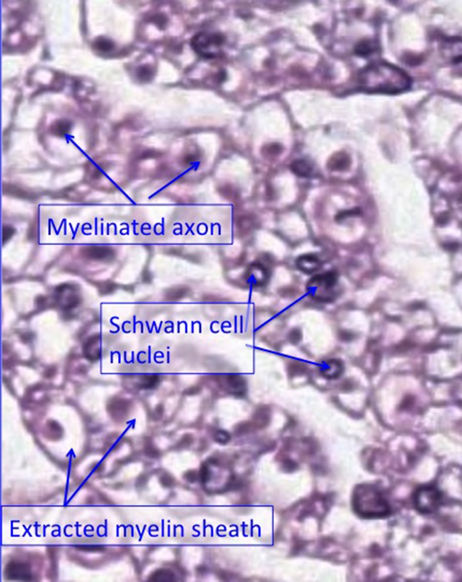
The endoneurium is not well stained in the preparation shown above, and is between the axons (which look like small circles). Organic solvents used in histological processing have extracted the lipid-rich myelin, so the myelin sheaths look empty. The dot in the middle of each circle is the axon. Remember that the only nuclei within the nerves represent the Schwann cells that produce the myelin or cells associated with blood vessels within the nerve.
All nerve fibers will have epineurium and endoneurium and depending on their size, may or may not have perineurium. Here is a cross section of a smaller nerve (below).

While epineurium is evident, no perineurium is present. The endoneurium cannot be distinguished from the Schwann cell cytoplasm surrounding the axons.
Below is a section of several nerves cut longitudinally.
Optional activity:You can practice looking at a cross-section of a peripheral nerve using a virtual microscope with guiding text in the right panel at this link.
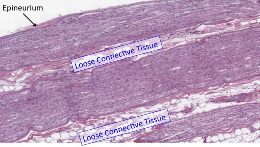
At higher magnification, most of the nuclei that you see here belong to Schwann cells (below).

The flat, wavy appearance of the cells/nuclei is characteristic of a peripheral nerve in longitudinal section. The axons and associated myelin appear as wavy lines.
Optional activity:You can practice looking at a longitudinal section of a peripheral nerve using a virtual microscope with guiding text in the right panel at this link.
Slide 2. Spinal dorsal root ganglion
Here is a spinal dorsal root ganglion that contains the neuronal cell bodies which provide afferent (sensory) information from the peripheral body (like the skin) to the spinal cord and brain (see below).
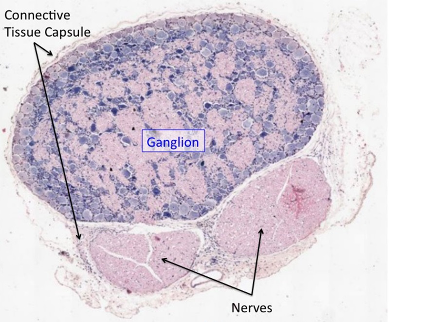
At low magnification you can see the connective tissue surrounding the ganglion, two profiles of adjacent nerves and the neurons and axons in the ganglion. The connective tissue around the ganglion becomes continuous with the epineurium and perineurium of the peripheral nerve. At higher magnification (see image below), note the large cell bodies of the neurons (asterisks) and the much smaller nuclei of the glial satellite cells (arrows).
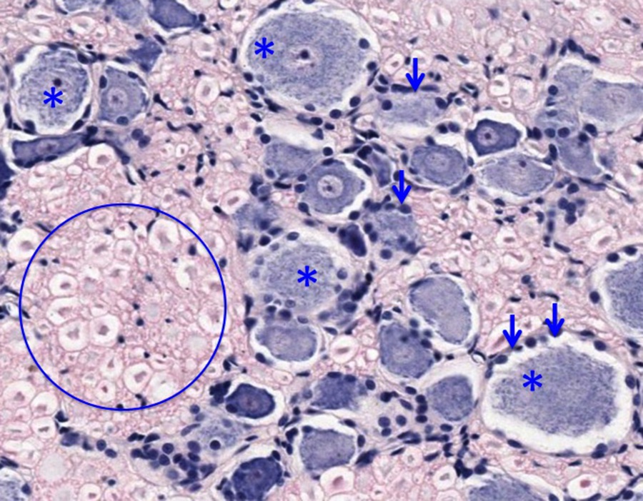
The cell bodies of the neurons contain lots of polyribosomes and rough endoplasmic reticulum (also known as Nissl substance) and hence are quite basophilic. Some neurons display nuclei with prominent nucleoli. The circle encloses multiple axons cut in cross section. The small, flattened nuclei that populate the nerve bundles are Schwann cells.
Although difficult to appreciate in histological sections, the neurons in the dorsal root ganglia are a special type. Called pseudo-unipolar, they have a single projection from the cell body that subsequently splits in two, one part projecting out to the periphery and the other to the dorsal horn of the spinal cord. Since these pseudo-unipolar neurons do not project dendrites to other adjacent neurons or receive input from other neurons within the ganglion, glial satellite cells can pack densely around the neuronal cell bodies, as seen below.
Optional activity:You can practice looking at a dorsal root ganglion using a virtual microscope with guiding text in the right panel at this link.
Slide 3. Normal Spinal Cord
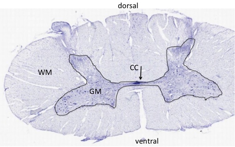
At low magnification, note the difference between the gray and white matter of the spinal cord (image above); the gray matter (GM) has many more large cell bodies than the white matter. The white matter (WM) surrounds the gray matter in the spinal cord, and has neuronal axons, but no neuronal cell bodies. The nuclei in the white matter are those of glial cells, astrocytes and oligodendrocytes. The white matter has both myelinated and unmyelinated axons. The axons are cut in cross section and hard to visualize even at high power, because most are traveling either up or down the cord. The organization of the gray matter resembles an H, with symmetrical dorsal and ventral horns. Note the large motor neurons in the ventral horn that send axons to the skeletal muscles of the body. The dorsal horn receives afferent (incoming) axons that bring sensory information from the periphery. As you saw in the previous sample, the cell bodies for these afferent sensory axons reside in the dorsal root ganglia and not in the spinal cord. In the very middle of the section, you can see the central canal (CC) as an elongated ring of small dark stained nuclei, the ependymal cells.
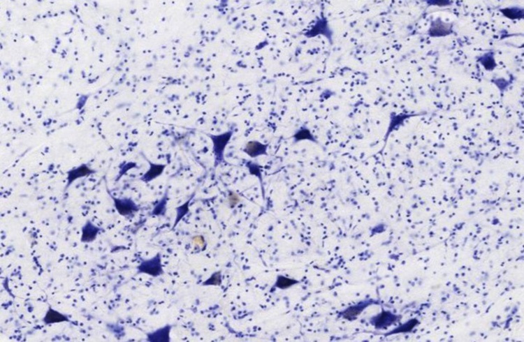
At higher magnification, note the basophilic pyramidal-shaped motor neuron cell bodies surrounded by nuclei of smaller glial cells (above).
Optional activity:You can practice looking at a normal spinal cord using a virtual microscope with guiding text in the right panel at this link.
Slide 4. Normal Cerebellum
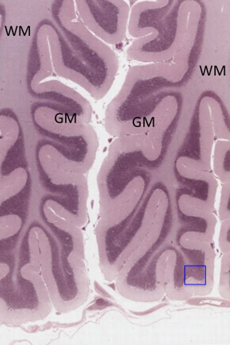
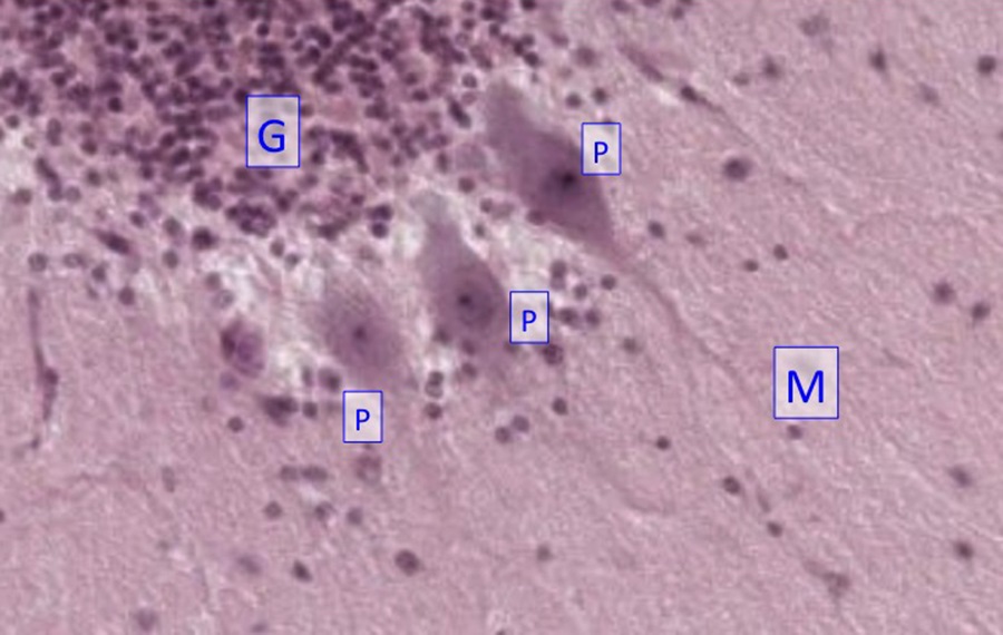
The section above is through the cerebellum, a part of the brain, is stained to show neuron and glial cell bodies, and proximal dendrites of neurons. The gray matter (GM) of the cerebellum is located superficially to a core of white matter (WM=axons). The inset shows a higher magnification view of the region boxed in the red square. The gray matter is organized into three layers:
- The molecular layer (M), a relatively cell-free layer close to the pial surface;
- A single layer of large Purkinje cells with globular cell bodies (P);
- The granule cell layer (G), a layer of small neurons just below the Purkinje cells.
Granule cells are among the smallest neurons. Purkinje cells are among the largest neurons in the brain. The white matter is composed of myelinated axons: the myelin is produced by oligodendrocytes.
Optional activity: You can practice looking at the cerebellum of the brain using a virtual microscope with guiding text in the right panel at this link.
Slide 5. Human cerebral cortex
At low magnification, you can see two gyri and a sulcus (S) dividing them in the section of the cerebral cortex below.
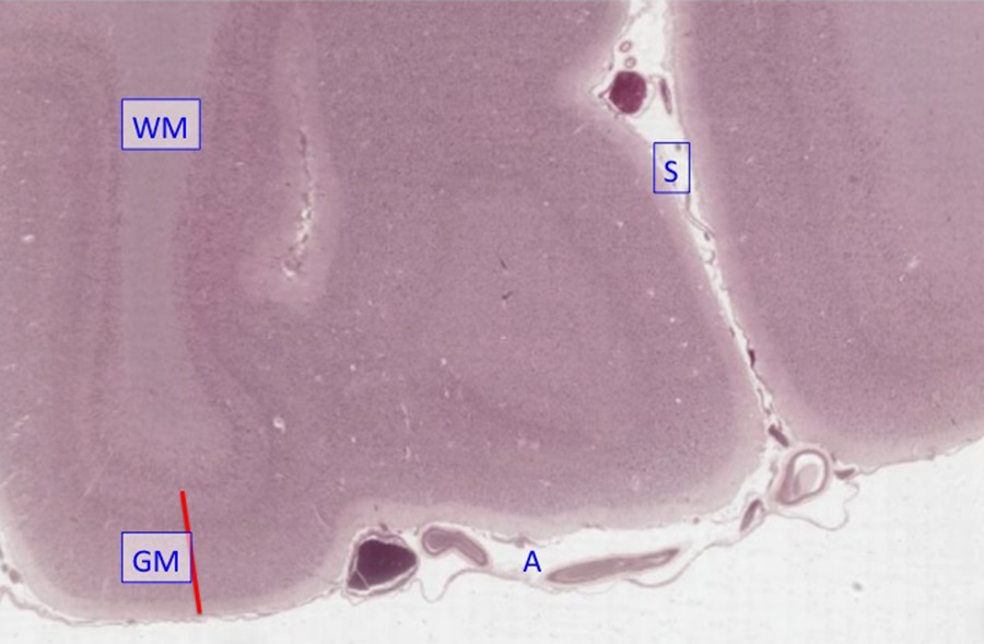
The human cerebral cortex is highly folded into these gyri and sulci. On the surface of the gyri and in the sulcus, you can see two layers of the meninges, the arachnoid (A) containing the blood vessels and the pia, which is cell layer immediately opposed to the neural tissue. The gray matter of the cortex (GM), has six layers of neurons and the white matter (WM) has small glial cells and axons of the cortical neurons, but no neuronal cell bodies. In the cerebral cortex, the gray matter is on the outside and the white matter is on the inside, the opposite of the spinal cord.
Slide 6. Cerebral cortex
Another section of the cerebral cortex with better preservation of the tissue:
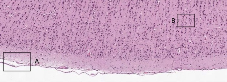
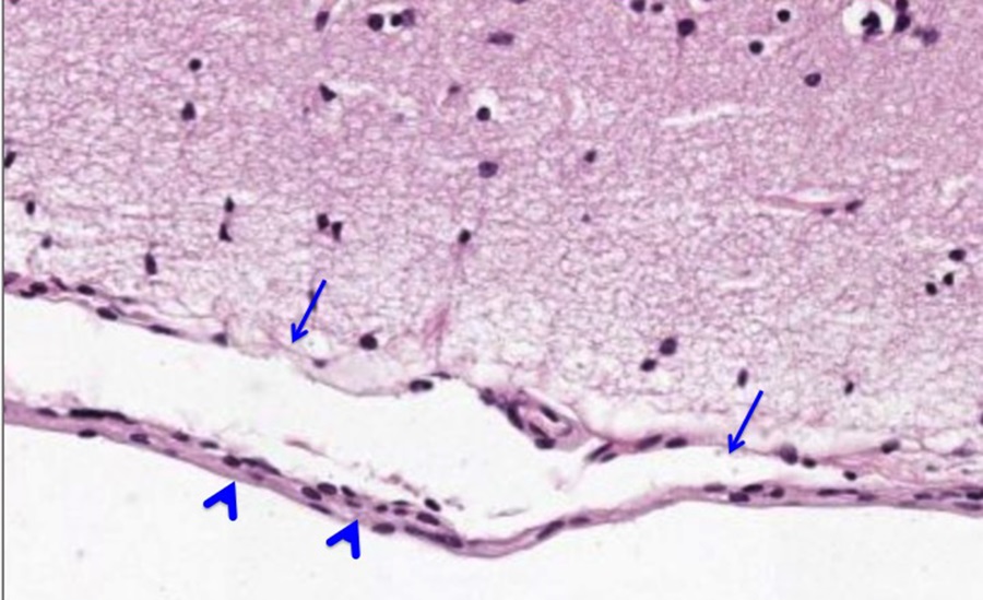
The area boxed in “A” is shown below of the meninges. Note the pia (arrows) and arachnoid (arrowheads):
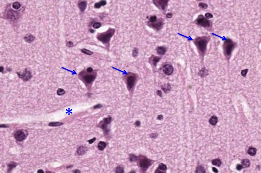
The region boxed in “B” is the inset below and shows the pyramidal neurons (arrows; named for the shape of their cell bodies) that are characteristic of the cerebral cortex and a small blood vessel (asterisk):
Slide 7. Golgi stained slide of rat cerebral cortex
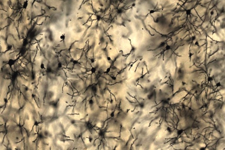
With this golgi stain (above), some neurons and some astrocytes are stained black and so the details of the cells can be resolved. Note that only a small number of the total cells are labeled with stain, which is characteristic of the Golgi stain. The basis for this seemingly random staining is not known.
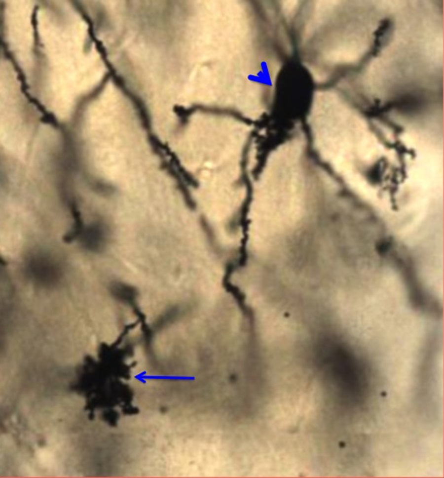
At higher magnification a protoplasmic astrocyte (arrow) and a pyramidal neuron (arrowhead) are shown below:
The image below shows spines on the apical dendrites of these pyramidal neurons (arrows):
Optional activity: You can practice looking at the neurons of the brain, specifically purkinje cells, using a virtual microscope with guiding text in the right panel at this link.
This Chapter’s PDF
- Note: The interactive features of this chapter are not reproducible in this PDF format.
ARCHIVAL PRE-CLASS MATERIALS FOR THIS SESSION:
- Pre-recorded videos: Nervous Tissue

Feedback/Errata