Histology of the Central Nervous System
SESSION OBJECTIVES
PRE-CLASS REQUIRED ASSIGNMENTS FOR THIS SESSION:
- Skim the section titles, bolded terms, and image captions from Junqueira’s Basic Histology, Chapter 9 (Click here for FMR library resources) to fill in any knowledge gaps you need.
A brief forward: This Pressbooks chapter both reviews and expands your neurohistology topic from the FMR Block. Many thanks to Drs. Jon Mallatt, PhD and Kate Mulligan, PhD for their significant contributions in rewriting and enhancing the original content.
NEURAL TISSUE OCCUPIES TWO MAIN DIVISIONS OF THE NERVOUS SYSTEM
I. The central nervous system (CNS) consists of the brain and spinal cord; most CNS cells are derived from the neural tube. This is the focus of today’s lecture.
II. The peripheral nervous system (PNS) consists of peripheral ganglia and nerves; most PNS cells are derived from the neural crest.
CNS DIVISIONS
The CNS can be divided into gray and white matter:
-
- Gray matter consists mostly of neuronal cell bodies, dendrites and axon terminals (together called neuropil), and glial cells. Examples of gray matter are the cerebral cortex and the dorsal and ventral horns of the spinal cord. In the brain, gray matter is organized into layers that form cortexes (e.g. cerebral and cerebellar cortexes) or into nuclei. A brain nucleus is a 3D cluster of neurons with a single function (e.g., the trigeminal motor nucleus contains neurons innervating muscles of mastication).
-
- White matter is made up of the axons of neurons whose cell bodies are located in the gray matter. Examples of white matter are the corpus callosum and the tracts of the spinal cord. White matter appears homogenous in gross material and in light microscopic preparations. However, careful gross dissection and modern imaging techniques (such as diffusion tensor imaging) demonstrate that bundles of axons (tracts) can intersect in complex ways, especially in the subcortical white matter of the brain.
MYELIN vs NISSL STAINS
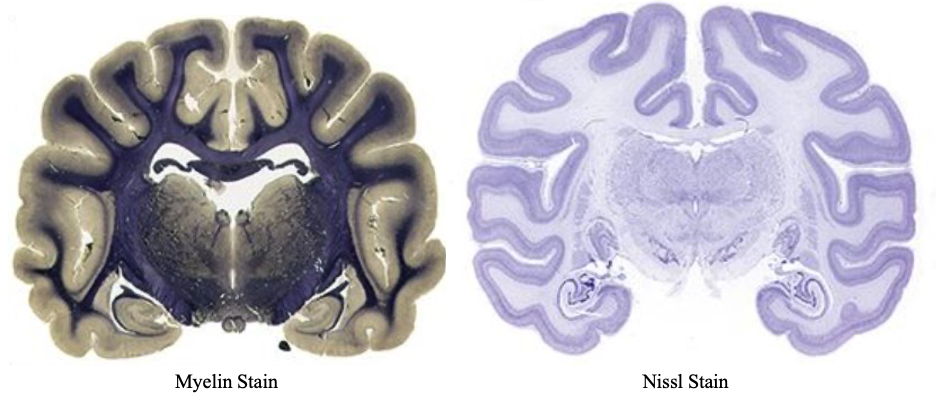
Myelin and Nissl stains offer complementary views that are used in teaching to differentiate between gray and white matter. Myelin stains react with myelin’s lipid so they stain the white matter very dark, while gray matter is pale. In contrast, Nissl stains are basic dyes that color nucleic acids blue or purple, thus staining nuclear chromatin, ribosomes, and RER. Consequently, they highlight cell bodies of individual neurons making gray matter stand out, while white matter is nearly unstained and appears light.
COMPARISON OF THE SPINAL CORD AND BRAIN
Everywhere in the CNS, gray matter lies deep—against the central canal of the spinal cord and the ventricles of the brain. In the spinal cord, this gray matter forms dorsal and ventral columns, which together form the shape of an H in transverse section. White matter (ascending and descending tracts) surrounds the gray matter. Specific white-matter tracts are located in the same relative area regardless of the level of the cord. For example, the dorsal columns contain the tracts carrying proprioceptive information, while tracts carrying pain and temperature are found in the anterolateral white matter.
The brain resembles the spinal cord in that deep gray matter likewise surrounds the ventricles and white matter lies external to these deep nuclei. However, there is a big difference: During brain development, some of the deep gray migrated out to form nuclei embedded in the white matter, and, in the cerebrum and cerebellum some gray migrated all the way out to the external surface to form the sheetlike cerebral cortex and cerebellar cortex.
CELL TYPES: There are two major cell types in the nervous system:
1. Neurons
2. Glia (supporting) cells
CNS NEURONS
Recall that neurons are electrically excitable cells that are highly polarized structurally. They communicate signals (often over great distances) by means of action potentials propagated along axons, and transmitted to other neurons and cells at special junctions called synapses.
Knowledge check: Identify all of the structures of a CNS neuron:
- Cell body/soma:
- Neuron cell bodies contain a large, round nucleus with a prominent nucleolus. The nucleus resembles an owl’s eye.
- Neurons actively synthesize proteins to maintain their long processes and to produce neurotransmitters. Their cell bodies therefore contain large amounts of rough ER and free ribosomes, in clusters called Nissl bodies or Nissl substance. Be aware that Nissl substance is not some distinct and unique organelle as was once thought, just large clusters of RER and ribosomes.
Knowledge check: Fill in the blank
- Dendrites
- Dendrites are processes, usually highly branched (dendrite=tree), that receive stimuli from other neurons at synapses and conduct these to the cell body.
- Most neurons in the CNS have many dendrites and are therefore called multipolar.
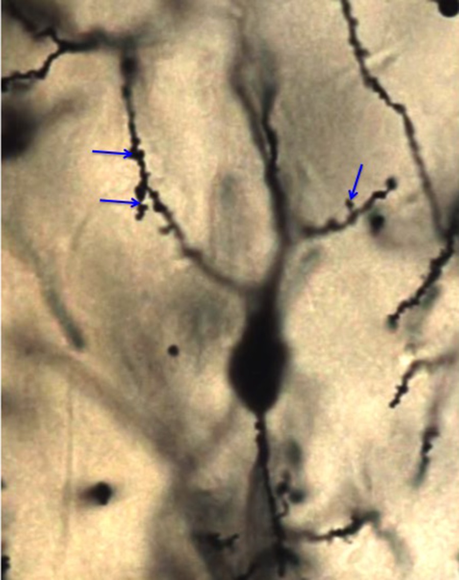
- In many neurons, dendrites are covered with spines, which are sites of synaptic contact (NOTE: synapses can also be on the non-spiny parts of dendrites).
- Spines: small protuberances from the dendritic surfaces that usually receive excitatory input. Their size and shape change over time and correlate with synaptic strength. New spines are made during learning and their retention correlates with the retention of specific memories.
- Dendritic cytoskeleton contains microtubules and associated proteins (MAPs). Neuron-specific beta tubulin is used to identify neurons by immunolabeling techniques. MAP2 stabilizes the microtubules in dendrites.
- Axons
- Most neurons possess a single axon that conducts action potentials away from the cell body.
- Axons arise from the cell body at the axon hillock, where action potentials are initiated.
- Axons are smooth processes (without spines) and usually do not branch near the cell body, but may give rise to infrequent collateral branches along their course.
- Often, axons branch profusely at their end at terminal arbors.
- Axon cytoskeleton has prominent microtubules and intermediate filaments made of neurofilament protein. Neurofilament protein binds silver, so silver nitrate stains for axons were classically used to describe axon pathways.
- Axoplasmic transport: Axons can be many centimeters and even meters in length. There are no ribosomes in axons, so no protein synthesis. To send cellular components along the axon from their point of synthesis in the cell body, neurons have a specialized transport system called axoplasmic transport, of two basic types:
- Fast transport-400 mm/day
- mostly for vesicles that move along microtubule tracks
- mediated by kinesin in the anterograde direction
- mediated by dynein in the retrograde direction
- microtubule-disrupting drugs cause neuropathy
- Slow transport-1 mm/day.
-
- moves cytoskeletal elements and the associated proteins (tau), and some cytosolic enzymes.
- limits the growth rate of regenerating axons
-
- Fast transport-400 mm/day
-
- Synapses
- If you wish to review chemical synapses, please check your materials from FMR Block. Recall the presynaptic and postsynaptic elements separated by a synaptic cleft, and the secretory process by which neurotransmitters are released from synaptic vesicles into this cleft.
- Important neurotransmitters in the central nervous system are glutamate, GABA, glycine, norepinephrine, dopamine, acetylcholine, and more.
MORPHOLOGICAL TYPES OF NEURONS IN THE CNS
There are many different morphological types of neurons in the CNS. While the vast majority are multipolar, there are huge variation in the configurations of the dendritic trees.
Two large and important neurons types are:
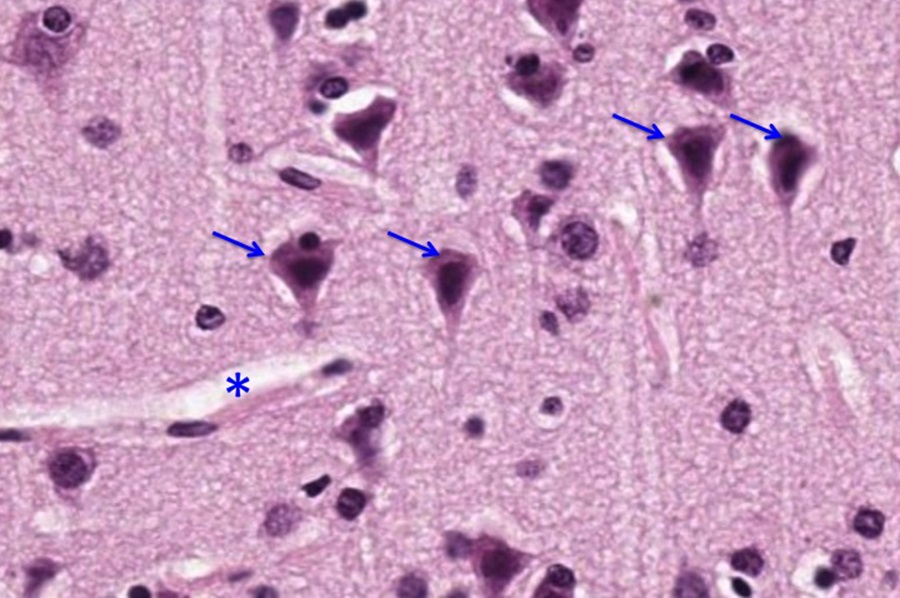
- Pyramidal cells of the cerebral cortex. These have a pyramidal cell body with an apical dendrite reaching to the cortical surface and a skirt of basal dendrites. Their axons project over long distances within the brain and spinal cord.*
- Purkinje cells of the cerebellar cortex. These are stunning neurons, as their cell bodies are tear-drop shaped and form a single layer in the cerebellar cortex, but the extremely dense dendritic tree is formed largely in 2 dimensions (almost flat).*
*More on these special neurons at the end of this Pressbooks chapter.
GLIA IN THE CENTRAL NERVOUS SYSTEM
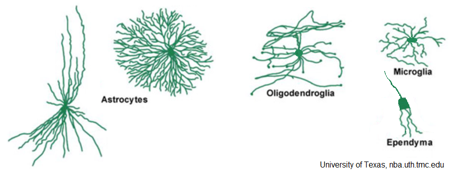
Glial cells, aka “neuroglia” (= “nerve glue”) occupy the space between neurons, separating neurons from each other and from blood vessels. Four types of neuroglial cells have been classically identified in the CNS: astrocytes, oligodendrocytes, microglia, and ependymal cells. All of these nourish and support neurons, while performing special functions of their own.
- ASTROCYTES
- The star-shaped astrocytes (astroglia) are not all the same and neuroscientists are just beginning to figure out how much diversity there is in these cells. However, traditionally they have been divided into two basic types: fibrous (in the white matter) and protoplasmic (in the gray matter). These are shown at left and right, respectively, in the figure above.
- Functions of astrocytes:
- Control blood flow to brain regions
- Initiate and maintain blood-brain barrier
- Modulate ionic environment of the fluid around neurons
- Aid synaptic function
- Send signals to neurons and glia
- Help neuro-repair
- Store glycogen to nourish neurons
- In astrocytes, the intermediate filaments of the cytoskeleton consist of “glial-fibrillary acidic protein” or GFAP. Astrocytes increase GFAP expression with damage or when nearby neurons are under stress and so this can be an early marker of neural damage.
- The most deadly brain tumors resemble astrocytes and are thought to derive from astrocytes. The most aggressive of these astrocytomas is glioblastoma multiforme.
- OLIGODENDROCYTES
- Oligodendrocytes (oligodendroglia) are most abundant in the white matter of the CNS, where they form the myelin sheaths of axons.
- The end part of each oligodendrocyte-process wraps around an axon in many concentric layers of plasmalemma to form a segment of myelin.
- Oligodendrocytes wrap only a short stretch of each axon, but they can wrap myelin around up to 50 axons.
- Recall that myelin, with its intervening nodes of Ranvier, increases the effective space constant of the axon membrane so that the action potential skips from node to node (saltatory propagation) and this greatly increases the conduction velocity.
- More recently discovered are satellite oligodendrocytes in the gray matter. They lie around neuron cell bodies, nourishing those and helping with neuronal signaling.
- EPENDYMAL CELLS
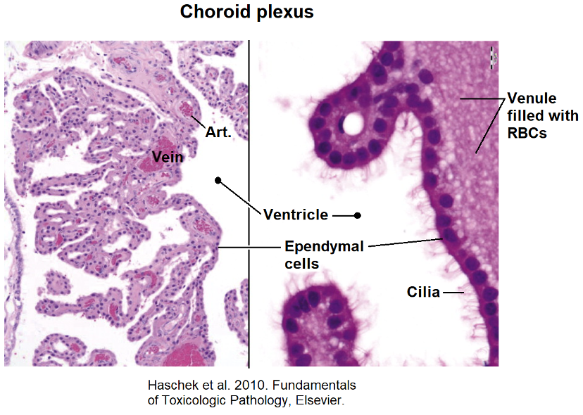
- Ependymal cells form an epithelium-like layer lining the ventricles of the brain and the central canal of the spinal cord. These cavities contain the cerebrospinal fluid (CSF).
- The apical surfaces of ependymal cells are covered with cilia, which help to circulate the CSF.
- In a few regions of the brain (mainly the hippocampus), specialized neural stem cells are mixed in with the ependymal cells. These stem cells have a single primary cilium and generate new neurons throughout your life. In the hippocampus, these new neurons are important for new memories. Despite this, the great majority of CNS regions cannot form new neurons.
-
- CHOROID PLEXUS
- Choroid plexus is a highly folded membrane on the roof of the brain ventricles, with its folds (“villi”) pushing down into some of the ventricles.
- It consists of ependyma overlain by a richly vascular connective tissue. It secretes the cerebrospinal fluid as a filtrate from its capillaries that is chemically modified by its ependyma.
- CHOROID PLEXUS
-
-
- The CSF acts as a cushion for the brain, suspending it in fluid within the dura. It also floats, and thus lightens, the central nervous system.
- A blood-CSF barrier is established by the zonula occludentes (tight junctions) between the choroid plexus ependymal cells.
-
CELLS NOT DERIVED FROM THE NEURAL TUBE RESIDING IN THE CNS
- MICROGLIA
- Microglia are small (hence “micro”), highly branched cells with processes covered with thorn-like projections.
- Microglia are migratory and phagocytic, the macrophages of the CNS. They are derived from circulating monocytes. They are the only neuroglial cells that do not develop (like neurons) from the neural tube.
BLOOD-BRAIN BARRIER
- The brain has a rich blood supply, but its capillaries have a restricted permeability that reduces exchange of large molecules between the blood and the brain cells.
- This is the blood-brain barrier, or BBB, and it stems from special tight junctions (zonulae occludentes) between capillary endothelial cells.
- Certain regions of the brain, like the hypothalamus, have a permeable BBB so that they can “sample” the blood for regulatory molecules.
- For further review, see the Nervous-Tissue topic back in FMR Block.
HISTOLOGICAL SECTIONS OF THE CENTRAL NERVOUS SYSTEM
By examining histological sections of different CNS regions, such as the spinal cord, cerebellum, and cerebral cortex, we can observe the intricate architecture that underpins their specific functions. Each region reveals a unique arrangement of neurons, glial cells, and axons, highlighted by various staining techniques that differentiate between the components.
Normal Spinal Cord
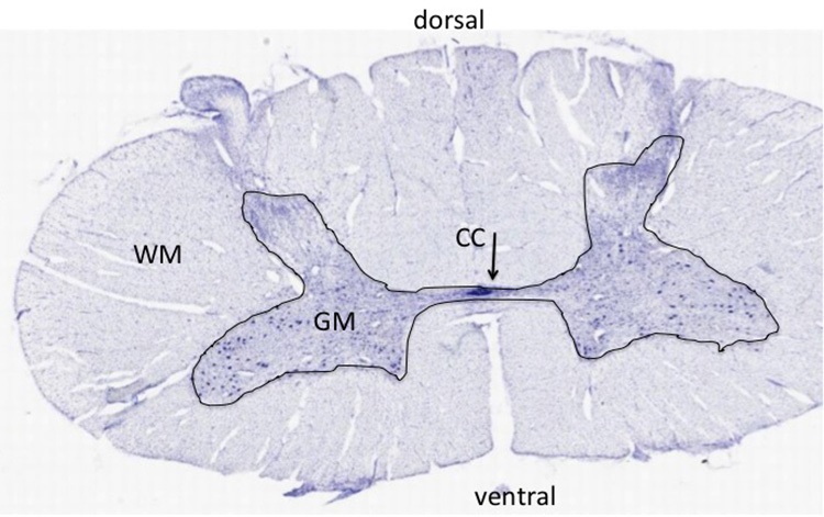
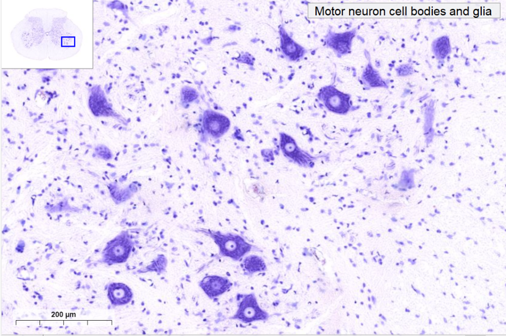
In this low-magnification, Nissl-stained section through the spinal cord, note the difference between the gray and white matter. The gray matter (GM) has the neuronal cell bodies. The white matter (WM) surrounds the gray matter in the spinal cord, and has axons but no neuronal cell bodies. The cell nuclei in the white matter are those of glial cells (astrocytes, oligodendrocytes, and microglia). The white matter has both myelinated and unmyelinated axons. The axons are cut in cross section as most are traveling up or down the cord. They are hard to visualize in Nissl-stained sections, which do not stain axons well. The organization of the gray matter resembles an H, with dorsal and ventral horns. Note the large motor neurons in the ventral horn that send axons to the skeletal muscles of the body. The dorsal horn receives afferent axons that bring sensory information from the periphery. The cell bodies for these sensory axons reside in the dorsal root ganglia and not in the spinal cord. In the very middle of the section, you can see the central canal (CC) as an elongated oval lined by small, darkly stained nuclei of ependymal cells.
At higher magnification, note the basophilic cell bodies of the large motor neurons surrounded by nuclei of smaller glial cells (below, from histologyguide.org).
Optional activity: You can practice looking at a normal spinal cord using a virtual microscope with guiding text in the right panel at this link.
Normal Cerebellum
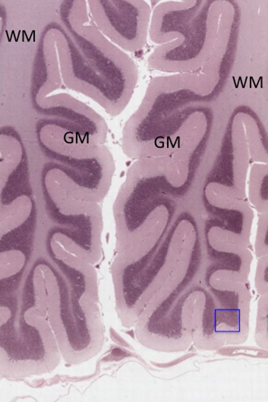
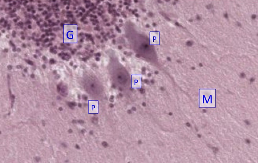
The section below through the cerebellum is stained to show neuronal and glial cell bodies, and proximal dendrites of neurons. The gray matter (GM) is located superficially to a core of white matter (WM=axons). The inset shows a higher magnification view of the region boxed in the blue square. The gray matter is organized into three layers:
- The molecular layer (M) is a relatively cell-poor layer close to the external, pial surface. It predominantly contains granule cell axons and Purkinje cell dendrites, with a few, scattered intrinsic neurons.
- A single layer of large Purkinje cells with globular cell bodies (P);
- The granule cell layer (G) of small, densely packed neurons just deep to the Purkinje cells.
Granule cells are among the smallest of all neurons. Purkinje cells are among the largest.
Optional activity: You can practice looking at the cerebellum by using a virtual microscope with guiding text in the right panel at this link.
Cerebral cortex
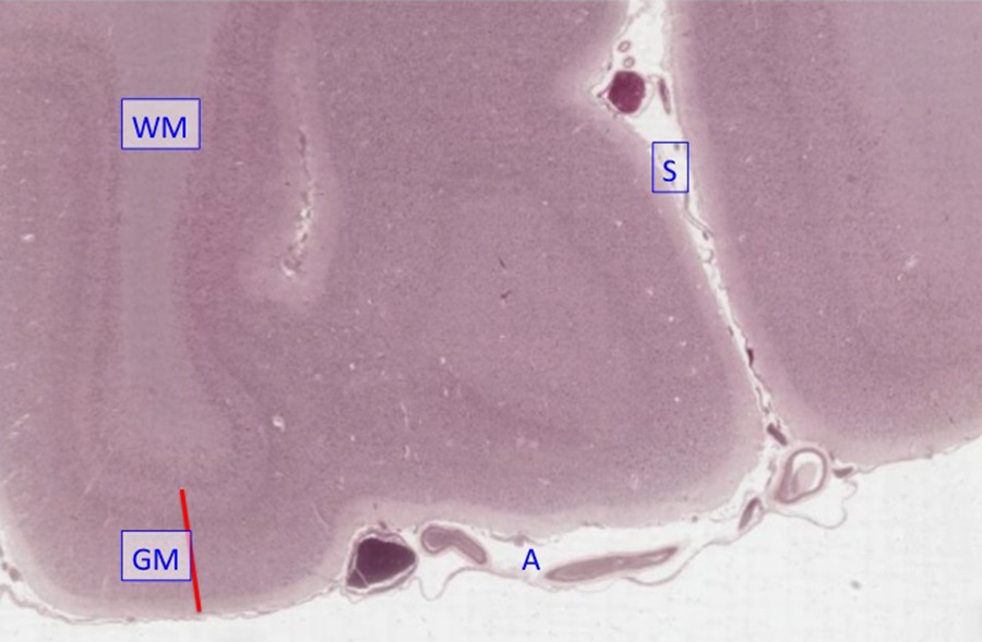
The human cerebral cortex is highly folded into gyri and sulci. The section below shows two gyri and a sulcus [S] between them. On the surface of the gyri ,you can see two layers of the meninges, the arachnoid (A) containing the blood vessels, and the pia, which is immediately opposed to the neural tissue. Pia continues into the sulci (S), but the arachnoid remains on the outer surface of the brain. The gray matter of the cortex (GM) has six layers of neurons, and the white matter (WM) has glial cells and axons of the cortical neurons, but no neuronal cell bodies. The cortex is an extra layer of gray, external to the white matter, and is not present in the spinal cord or brainstem
Another section of the cerebral cortex with better preservation of the tissue:
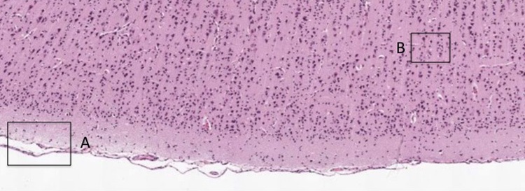
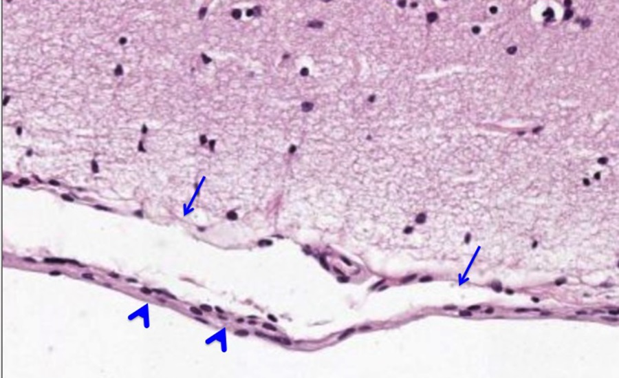
The area boxed at “A” is shown below and contains meninges. Note the pia (arrows) and arachnoid (arrowheads). Next figure will show the area in “B.”
The area boxed at “B” is shown below. Note pyramidal neurons (arrows; named for the shape of their cell bodies) that are characteristic of the cerebral cortex, and a small blood vessel (asterisk). The apical dendrites of pyramidal cells are oriented towards the pia.
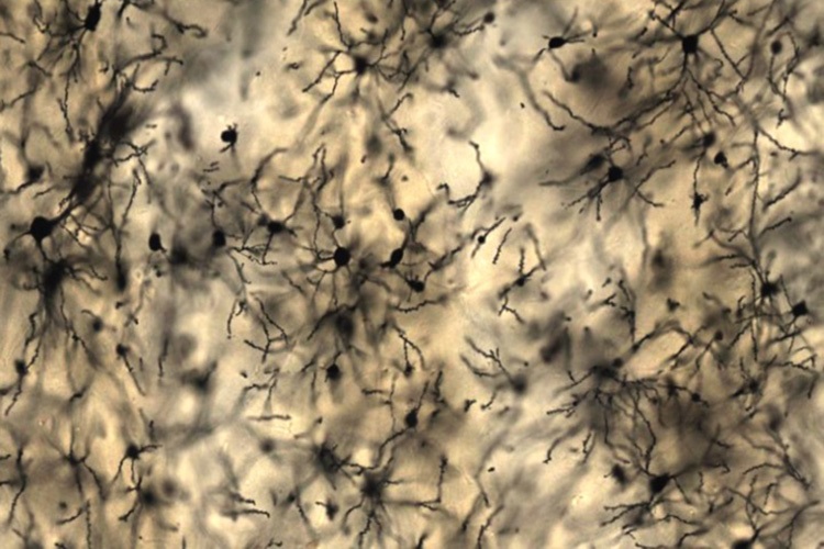
Golgi-stained slide of rat cerebral cortex
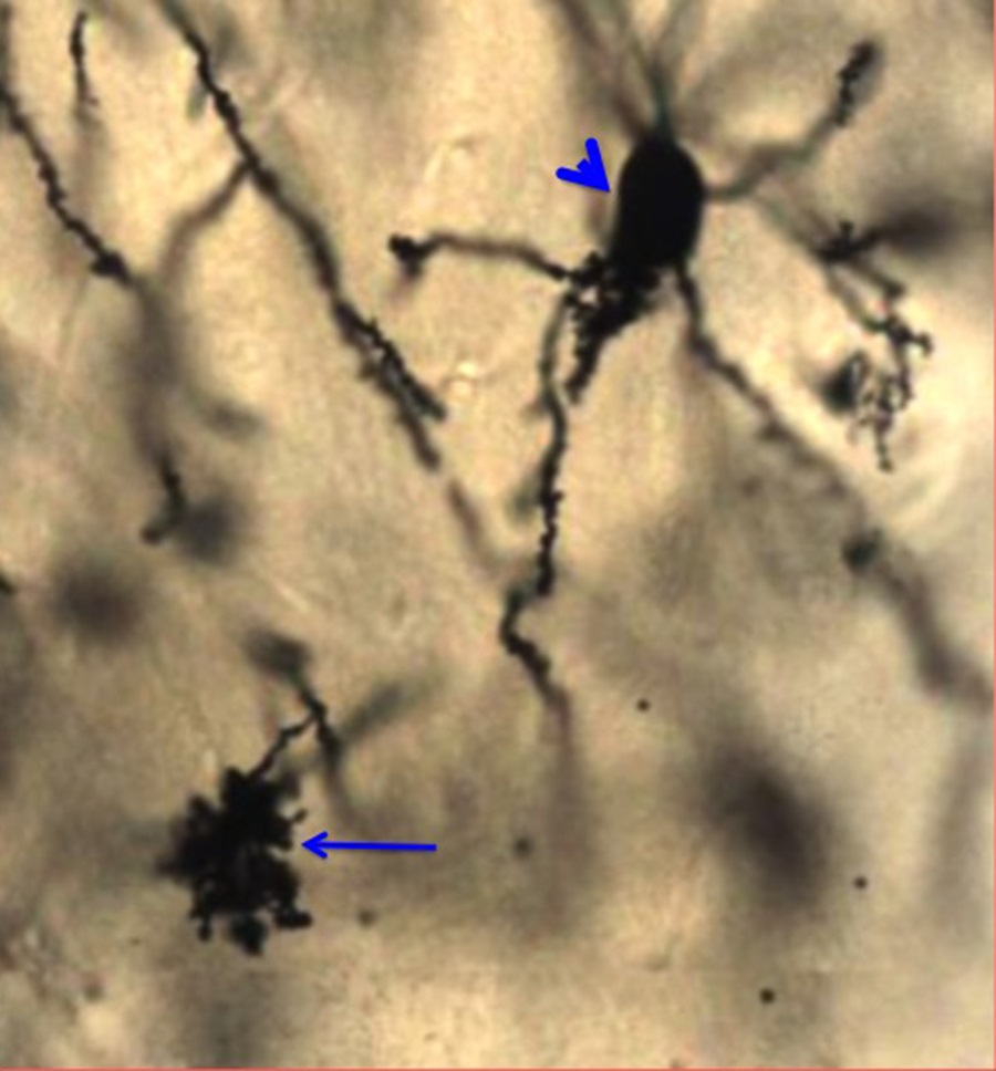
With this Golgi stain, some neurons and some astrocytes are stained black so the shapes of entire cells can be resolved. Note that only a small number of the total cells are stained, which is characteristic of the Golgi stain. The basis for this seemingly random staining is not known.
Below is a higher-magnification image of a pyramidal neuron (arrowhead) and a protoplasmic astrocyte (arrow):
The image below shows spines on the apical dendrites of pyramidal neurons (arrows):
Optional activity: You can practice looking at the neurons of the brain, especially pyramidal and Purkinje cells, using a virtual microscope with guiding text in the right panel at this link.
Knowledge check:
This Chapter’s PDF
- Note: The interactive features of this chapter are not reproducible in this PDF format.
ARCHIVAL PRE-CLASS MATERIALS FOR THIS SESSION:
- Pre-recorded videos: Nervous Tissue

Feedback/Errata