
2 Anatomy of the Large Vessels
Overview of the vessels
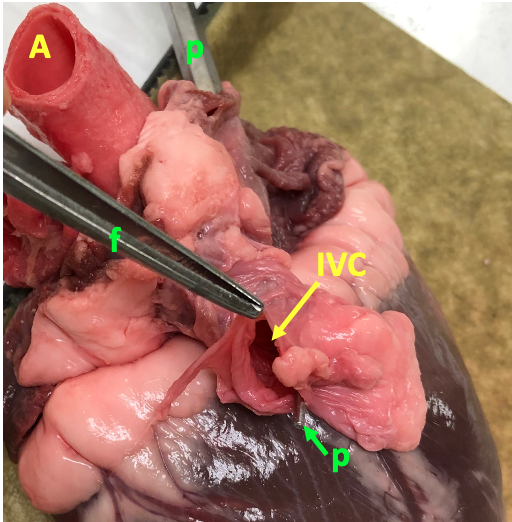
The arteries are the vessels that carry blood away from the heart. The veins are the vessels that return blood to the heart. As we shall see when we examine the histology of blood vessels, the arteries and the veins have the same basic three-layered structure. However, because arteries experience much higher pressures, they are tougher and thicker-walled. By contrast, veins are thin-walled and compliant, meaning they are stretchy and pliable. This distinction is illustrated in the photo below, which shows the aorta, the largest artery in the systemic circulation, and the inferior vena cava, the largest vein of the systemic circulation that returns blood to the heart from the lower body. The aorta is thick-walled and maintains its shape as a tube. The inferior vena cava is thin-walled and floppy like a deflated balloon.
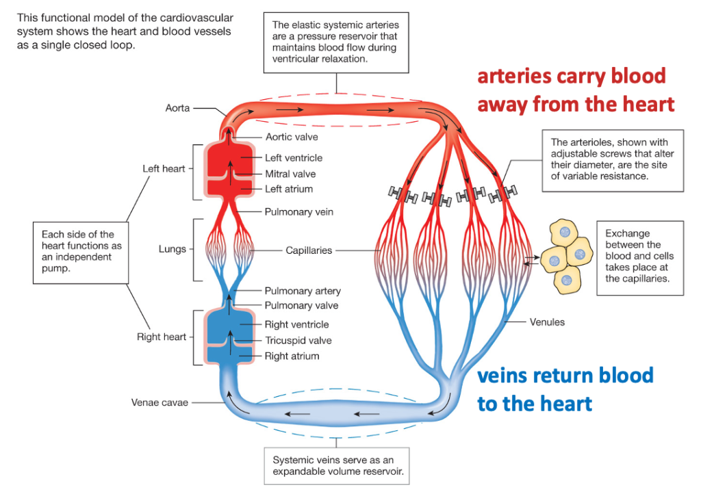
The figure below provides a nice overview of the heart and vessels of the circulatory system that you can use as a reference.
We will restrict our study to the large arteries and veins in the chest, and the major vessels of the coronary circulation that serves cardiac tissue. We will also learn the arteries in the abdomen and groin in order to understand the path used for transcatheter aortic valve replacement. The material to study includes videos from Acland’s Video Atlas of Anatomy, and pictures of a heart model from the Health Sciences Library.
Arteries
Aortic arch and its branches
The model of the heart provides a good starting point for seeing the major arteries adjacent to the heart. The letters in the figure are referred to in the text.
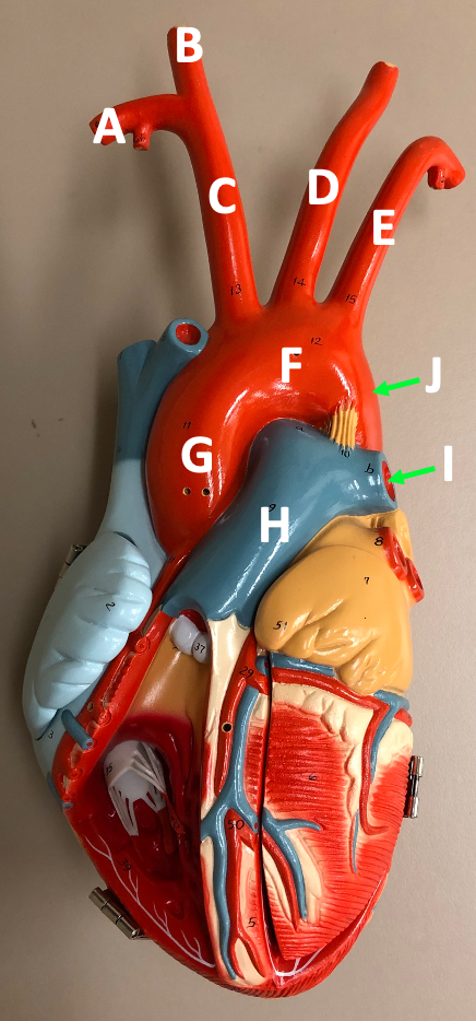
The large arteries that carry blood away from the heart emanate from the base of the heart. The pulmonary trunk (H) is slightly anterior of the ascending aorta (G)and pointing to the left. The pulmonary trunk divides into two pulmonary arteries (I indicates left pulmonary artery) that deliver blood to each of the lungs. The aorta heads superiorly (ascending aorta, G), tilting towards the right, before arching to the left (aortic arch, F). The aorta then descends through the thoracic and abdominal cavities. J shows the descending thoracic aorta.
Three large arteries that supply the arms and head are attached to the aortic arch: the brachiocephalic artery (C), the left common carotid artery (D), and the left subclavian artery (E). On the right, the brachiocephalic artery heads superiorly, then branches into the right subclavian artery (A) and the right common carotid artery (B). The subclavian artery is so named because it is deep to the clavicle. This artery supplies blood to the arms. The common carotid artery (which supplies blood to the head) travels superiorly in the neck, eventually branching to give rise to the external and internal carotid arteries. Where it branches, there is a swelling called the carotid sinus. This is a location for the carotid baroreceptors, sensory neurons that monitor the blood pressure.
There is no brachiocephalic artery on the left side. Instead, the left common carotid artery (D) and the left subclavian artery (E) directly branch off of the aortic arch. Both vessels head superiorly away from the arch, but the subclavian artery turns laterally just below the clavicle.
The Acland’s videos nicely show how these vessels appear in an actual human body. The first video focuses on the aorta and its major branches.
3.1.0 Ascending aorta, aortic arch and its branches
The figure below is a screenshot showing these major arteries.
acland’s arteries-top
Abdominal aorta and its branches
After the aortic arch, the aorta descends through the chest cavity (descending thoracic aorta), then enters the abdomen (abdominal aorta), bringing blood to arteries that supply the abdominal organs and the legs. At the level of the 4th lumbar vertebra, the aorta divides, forming the two common iliac arteries. The common iliac artery divides to give rise to the internal iliac artery (traveling medially into the pelvic cavity) and the external iliac artery, which supplies blood to the legs. The artery passes under the inguinal ligament and exits the abdominal cavity to lie in a position at the top of the thigh. After, it exits the abdomen, the artery is called the femoral artery. The femoral artery is a convenient access point for percutaneous (through the skin) cardiac valve surgery.
The figure below is an illustration of the abdominal aorta and its branches that you can use for reference.
figure from grays
There are two videos showing the large arteries that arise from the abdominal aorta. Watch Arteries of the hip region only up until the 1:06 minute mark.
2.1.14 Arteries of the hip region
Watch Arteries of the abdominal region from the beginning through “bifurcation of the aorta”, then jump to “common iliac artery” using the links on the left-hand menu.
3.3.14 Arteries of the abdominal region
Some more text with key screenshots
Veins
The superior vena cava and the inferior vena cava deliver blood directly to the right atrium, and are the largest veins in the body. The inferior vena cava is mainly located in the abdominal cavity. We will focus on the veins that bring blood to the superior vena cava. The vein that drains blood from the arms is called the subclavian vein, which runs parallel to the subclavian artery. The large vein that drains blood from the head is the internal jugular vein. This vein runs adjacent to the common carotid artery in the neck. The subclavian and internal jugular veins join to form the brachiocephalic veins. Because the superior vena cava lies on the right side of the body, the right brachiocephalic vein is short and vertically oriented, while the left brachiocephalic vein is longer and obliquely oriented.