
1 Anatomy of the Heart
The heart lies within the pericardium
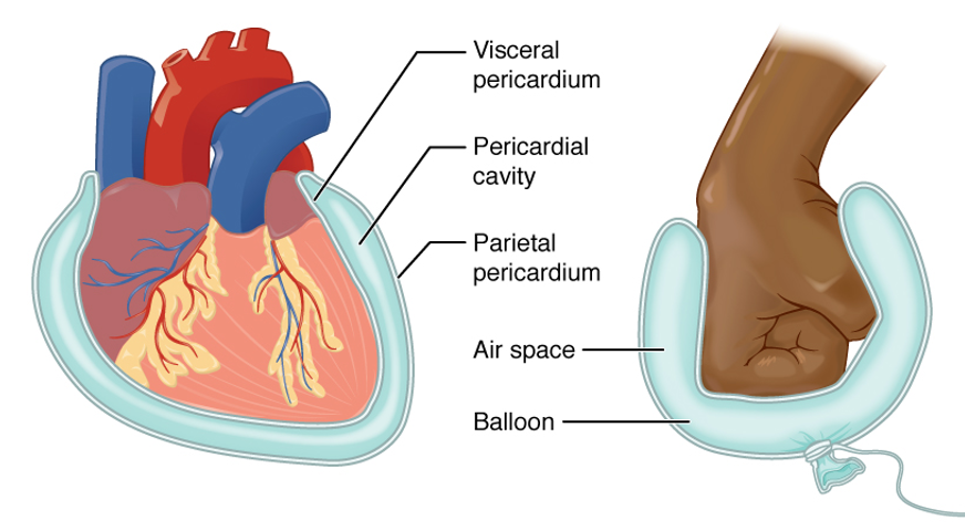
The heart is contained within a tough membranous sac known as the pericardium. As shown in the figure below, the pericardium is two layers: a visceral pericardium next to the heart and a parietal pericardium next to the surrounding tissues. The pericardial cavity is filled with fluid that lubricates the beating heart and reduces friction.
https://openstax.org/books/anatomy-and-physiology/pages/1-6-anatomical-terminology#fig-ch01_06_06
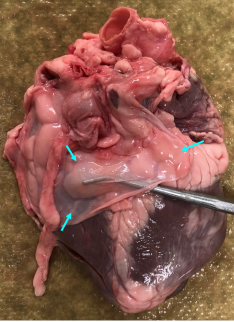
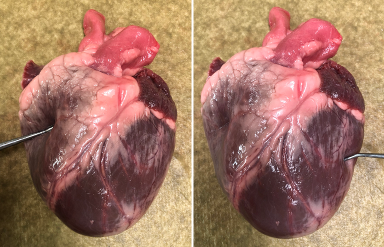
The figure below shows the posterior side of an isolated pig heart with a small amount of pericardium still attached. A probe can be seen through the thin layer of pericardium.
Optional, but highly recommended video
The video below, from Acland’s Video Atlas of Anatomy, nicely illustrates the pericardium. It also shows how the heart is situated in the body, and gives you a preview of the anatomy of the large vessels associated with the heart.
5.1.9 Pericardial sac, great vessels
External features of the heart
Below is an illustration showing the external features of the heart, that you can use for reference.
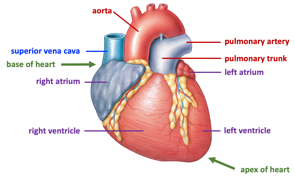
Note that when you look at the above figure or the picture of the heart below, it is displayed as you would see it in the body. Thus, the right side of the heart will be on your left, and the left side of the heart will be on your right. This is the general convention used in anatomical illustrations.
The picture below shows an anterior view of the dissected pig heart, with the pericardium removed. The letters indicated in the text refer to the labels on the picture.
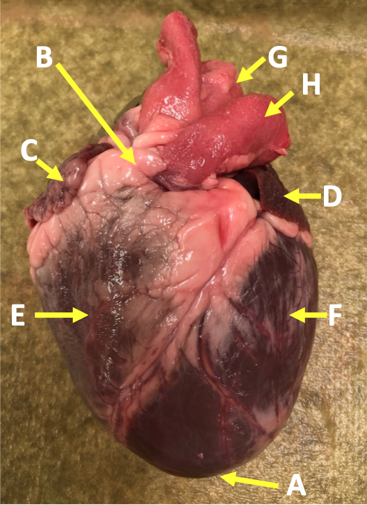
The inferior part of the heart is called the apex (A) because it comes to a point, like the apex of a cone. The superior part of the heart is referred to as the base (B). The major vessels of the heart are found at the base of the heart, along with the upper chambers, the right atrium (C) and left atrium (D). The atria are collapsed, but in a functioning heart, they would be stretched full of blood.
The anterior surface of the heart is characterized by the presence of the large arteries leaving the base of the heart, the pulmonary trunk (H) and the aorta (G). The pulmonary trunk is the vessel that divides to give rise to the two pulmonary arteries that deliver blood to each lung. The pulmonary trunk is somewhat anterior to the aorta, and although it is connected to the right ventricle, it tilts leftward. The aorta is slightly posterior to the pulmonary trunk and bound to it by connective tissue.
The majority of the heart tissue consists of the ventricles. The left ventricle (F) is stiff and solid because it is very thick-walled. By contrast, the right ventricle (E) has a much thinner outer wall. This difference is illustrated in the pair of photos below.
The right ventricle only needs to pump blood to the lungs, which lie close to the heart; by contrast, the left ventricle needs to pump blood throughout the body. Consequently, the pressures generated by the two sides of the heart are quite different, with average pulmonary arterial pressure being about one sixth of the systemic mean arterial pressure (MAP; average blood pressure).
Internal features of the heart
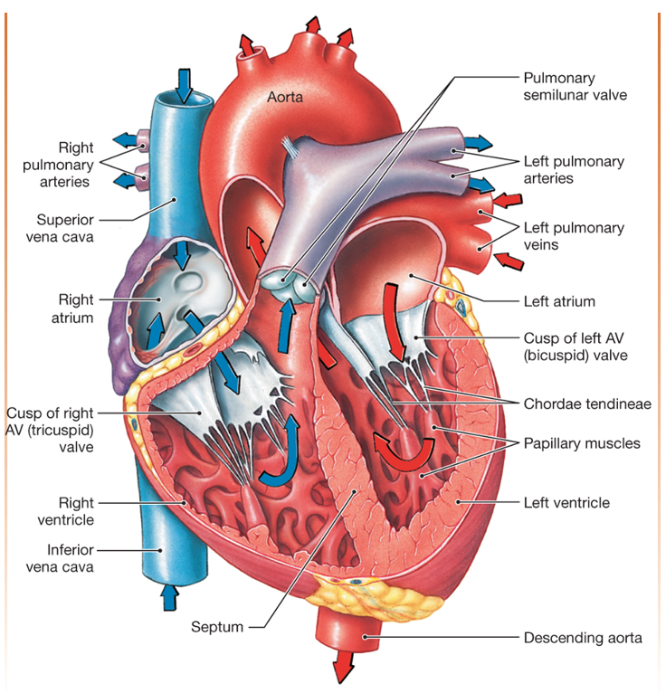
An internal view of the heart shows the interventricular septum that divides the two ventricles, as well as the valves and their associated structures. Below is an illustration showing the internal features of the heart that you can use for reference. This figure includes blue and red arrows that indicate the flow pattern of deoxygenated (blue) and oxygenated (red) blood.
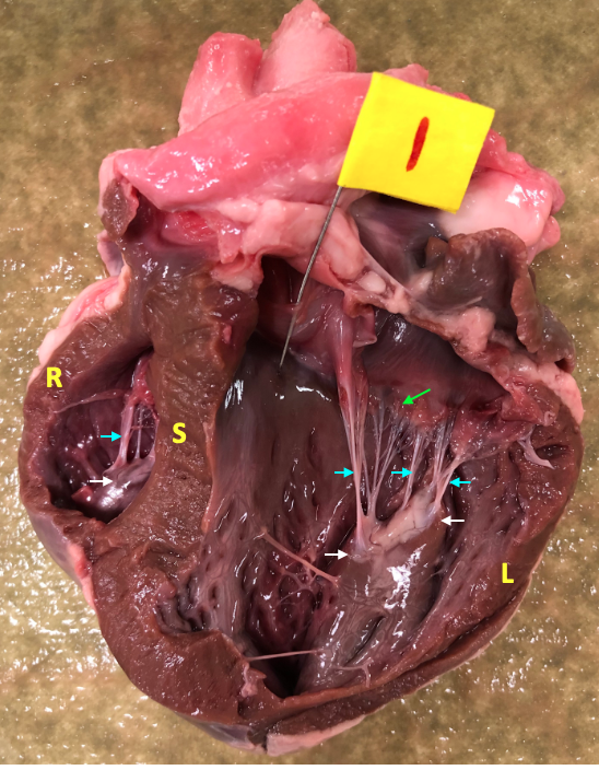
The figure below shows a similarly sectioned pig heart.
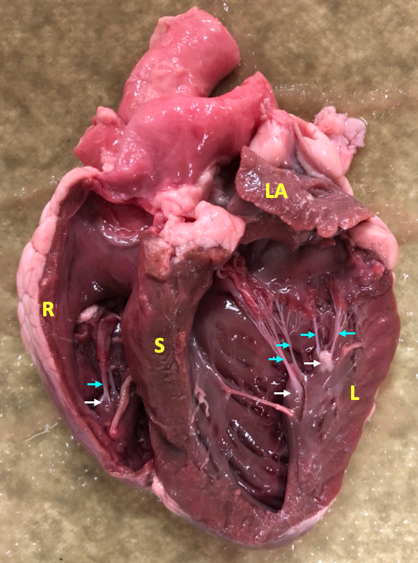
The left and right ventricles are separated by the large interventricular septum (S). Note the relative thickness of the walls of the right ventricle (R) and left ventricle (L). The inner surface of the heart is covered with irregular bands of tissue known as trabeculae carneae (these are visible in the lower part of the left ventricle in the above picture). The papillary muscles (white arrows) are distinct little hills of muscle that poke up from the inner surface of the heart. At the tips of the papillary muscles are the chordae tendineae (cyan arrows), strings of connective tissue that attach to the edges of the atrioventricular valves (AV valves).
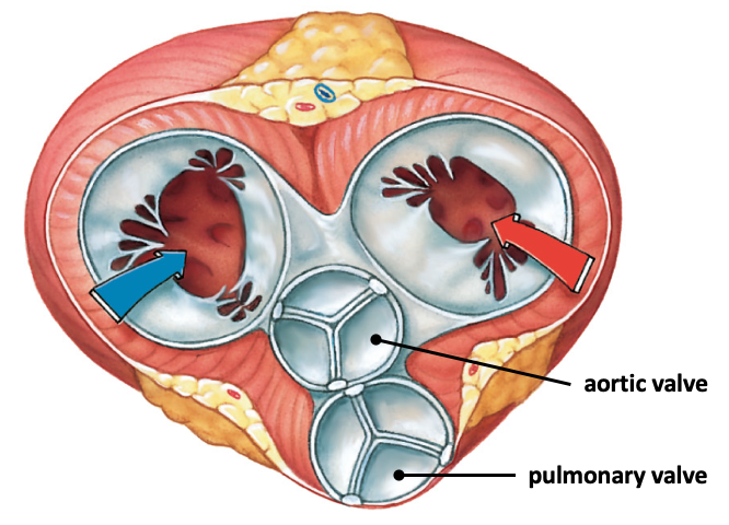
The heart contains four important valves that ensure that blood flows in one direction. The right AV valve (tricuspid valve) and the left AV valve (mitral valve) lie between the atria and ventricles, and prevent blood from flowing back into the atria during systole (systole is the term for the part of the cardiac cycle when the ventricles are contracting). The pulmonary valve and the aortic valve lie at the beginning of the large outflow vessels at the base of the heart, and prevent blood from falling back into the heart during diastole (diastole is the term for the part of the cardiac cycle when the ventricles are relaxed).
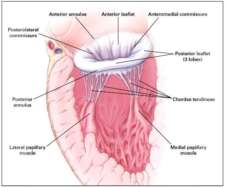
The valves consist of two or three cusps (also called leaflets) of delicate connective tissue that come together to block the flow of blood. The valve cusps of the AV valves look like torn curtains, and are easily identified because they have the chordae tendineae attached to them. The structure of the left AV valve is shown in the figure below.
The papillary muscles contract early during systole. This holds the valve cusps of the AV valves in place and prevents them from prolapsing.
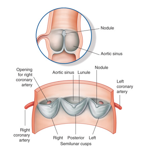
The aortic and pulmonary valves each have three valve cusps that look like little pockets or half-moons. For this reason, these valves are referred to collectively as the semilunar valves.
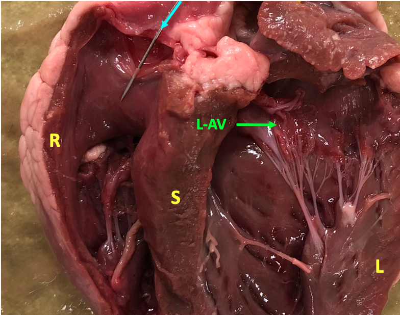
Below is a photo of the dissected heart, focussing in on the valves. One cusp of the pulmonary valve is pierced by a pin and the left AV valve is indicated by an arrow.
The figure below shows a different pig heart. This heart is dissected to show the aortic valve, which is indicated by the pin with the flag.
All four heart valves are located at the same plane in the heart and are attached to a fibrous layer of connective tissue called the cardiac skeleton.
The cardiac skeleton provides mechanical support for the valves. Since it is connective tissue, and not cardiac muscle, the cardiac skeleton also electrically isolates the atria from the ventricles. This is important for activating contraction of the heart in the appropriate sequence.
The action of the valves is nicely illustrated in the video from Acland’s Video Atlas of Anatomy linked below. When you click on the link, the video will open in a new tab.
5.1.6 Ventricles: outflow pathways
The action of the valves is demonstrated by pumping water through a dissected heart. In the video, you should be able to identify the following structures: aorta, aortic valve, pulmonary trunk, pulmonary valve, coronary artery, AV valve, chordae tendineae, papillary muscle.