6 Ventricles, Cerebrospinal Fluid, and the Blood-Brain Barrier
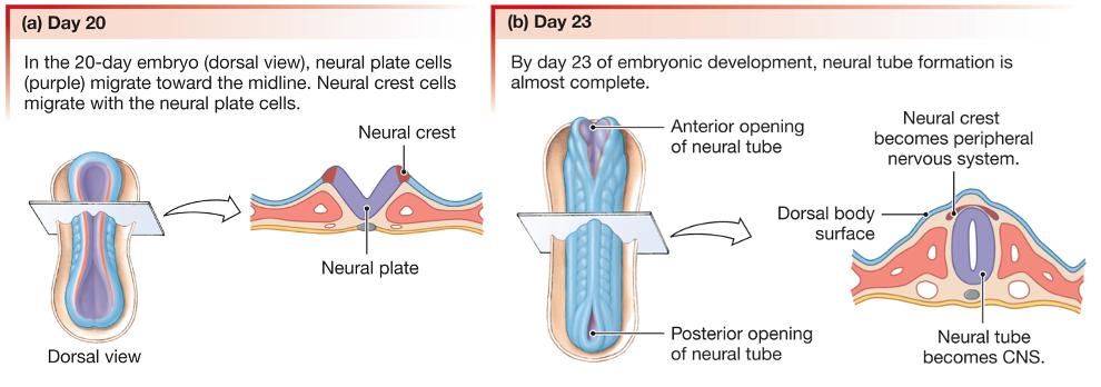
The central nervous system (CNS; brain and spinal cord) contains hollow spaces within it: the ventricles within the brain, and the central canal within the spinal cord. These spaces arise because the structures that will become the CNS develop from a tube. As the figure shows, early in development, the edges of the neural plate migrate toward each other to form the neural tube.
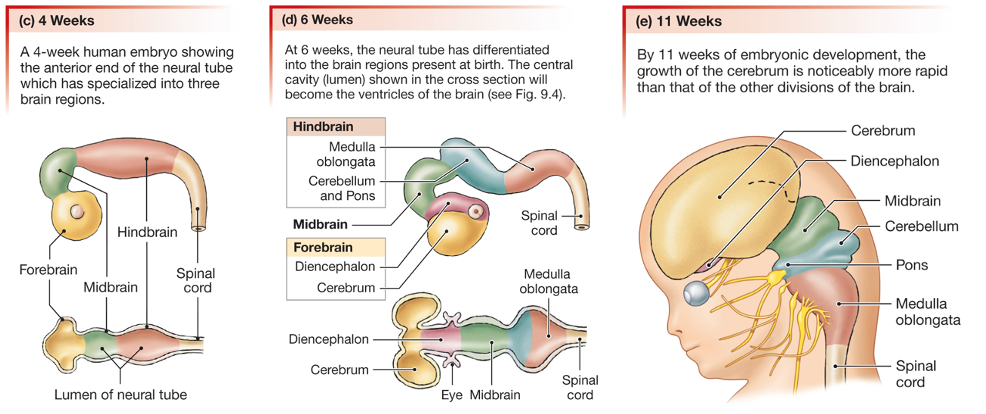
The neural tube undergoes regional specialization along its anterior-posterior axis. The anterior-most tissue expands greatly: this will become the cerebrum. The region just posterior to the developing cerebrum gives rise to the diencephalon, which includes the thalamus and the hypothalamus. The optic cup that will give rise to the retina in the eye also develops from the region. The midbrain, hindbrain (which includes the pons, cerebellum, and medulla oblongata) and the spinal cord develop from more posterior regions of the neural tube.
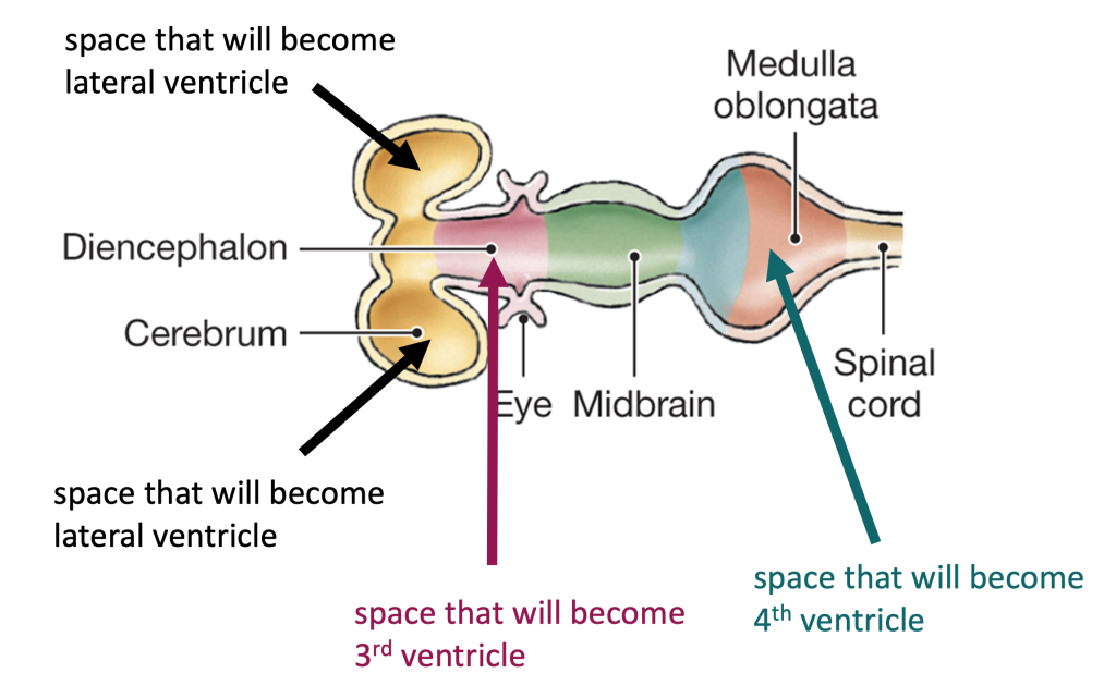
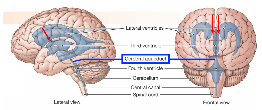
There are four ventricles: two lateral ventricles, a third ventricle, and a fourth ventricle. The figure below illustrates how the spaces in the neural tube will give rise to the four ventricles.
The next figure shows the final form of the ventricles in the brain. Each lateral ventricle is connected to the third ventricle by a passageway called the interventricular foramen (note the spelling: “inter-” meaning “between”, not “intra-“, which means “within”). The third ventricle is a very narrow space whose walls are made by the tissue of the diencephalon (thalamus and hypothalamus). The third ventricle is connected to the fourth ventricle by the cerebral aqueduct, a passageway that travels through the tissue of the midbrain.
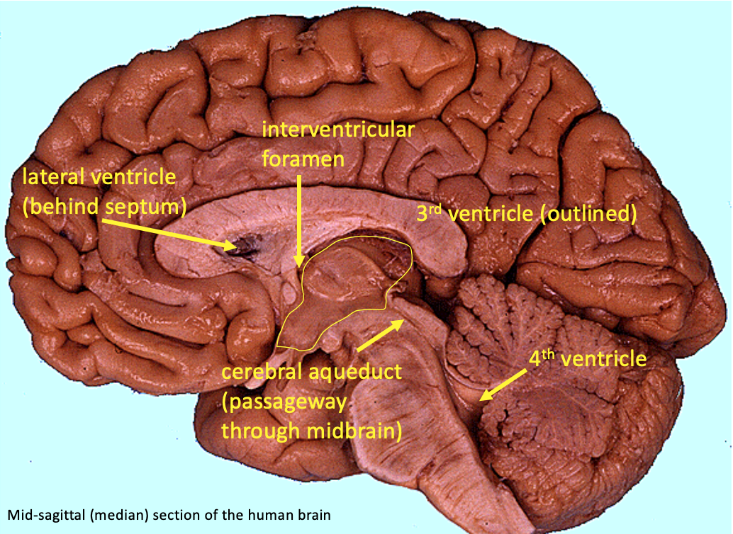
The figure below shows a mid-sagittal section (also called a median section) of a human brain, indicating the location of the ventricles and passageways.
The ventricles are filled with a fluid called the cerebrospinal fluid (CSF). CSF is produced at the choroid plexus, capillary-rich tissue found on the floor of the lateral ventricles; and the ceiling of the third and fourth ventricles.
The interstitial fluid of the CNS is isolated from the blood plasma by the functional barrier known as the blood-brain barrier. Endothelial cells in CNS capillaries are linked by tight junctions; thus, substances can only enter the interstitial fluid if they are transported across the plasma membranes of the endothelial cells.
What about the CSF? It can exchange with the interstitial fluid in the brain, but it does so minimally. CSF moves by bulk flow through the ventricles, exiting at particular passages into the subarachnoid space, a fluid-filled space which surrounds and cushions the brain and spinal cord. CSF drains back into the circulation at large veins in the skull via specializations called arachnoid villi.
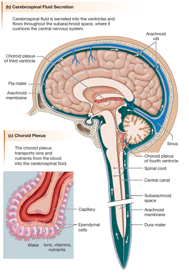
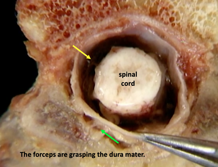
The subarachnoid space is created by meninges, connective tissue membranes that surround the CNS. There are three meningeal layers: a tough, thick outer layer called the dura mater, a middle layer called the arachnoid mater, and a delicate inner layer on the surface of the tissue called the pia mater. As shown in the figure below, the dura mater is attached to bones inside the skull. The arachnoid mater is located just inside the dura mater but it extends cobweb-like processes down to the pia mater. The fluid-filled space lies under both the arachnoid and dura and so is called the subarachnoid space.
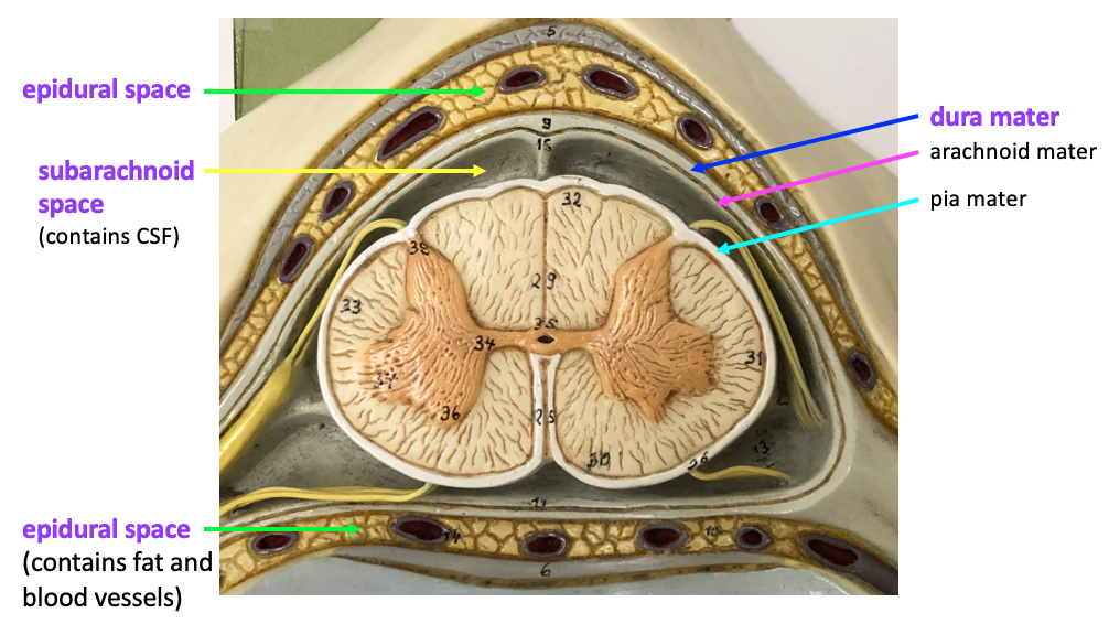
In the spinal column, the dura mater is separated from the vertebrae by a layer of fat tissue. This region, inside the bone but outside the dura mater, is called the epidural space. The epidural space is a location where local anesthetic can be injected. Local anesthetics are sodium channel blockers that can diffuse through the tissue to block action potentials in the nearby nerve roots.
The figure shows a model that is a cross-section of the spinal cord at the cervical level, with the meninges and spaces labeled.
Although all three meningeal layers are shown on the model above, in practice the arachnoid mater and pia mater are too delicate to see with the naked eye. The linked video from the Acland’s Video Atlas of Anatomy, shows the dura mater, subarachnoid space, and epidural space associated with the spinal cord. When you click on the link, it will open in a new tab.
3.1.8 Spinal cord (cross-section), spinal meninges, dural sac
For the purpose of this class, you need only to focus on identifying the structures described in the first half of the video (up to 1:22). These structures are labeled in the screenshot below.