20 Urinary System Histology
Structure of the Nephron
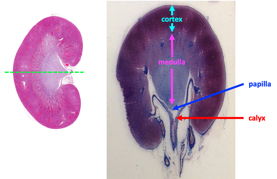
The nephron is the functional unit of the kidney. Each kidney contains roughly one million nephrons. The illustration on the left in the figure below depicts the kidney as it would look if sectioned along its long axis and shows how the nephrons are arranged between the two major regions of the kidney, the cortex and the medulla.
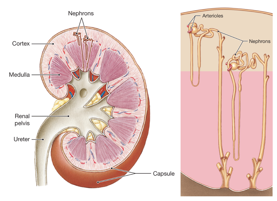
The figure on the right shows an enlarged view of two nephrons. We will focus on the structure and function of juxtamedullary nephrons (shown on the right). Juxtamedullary nephrons have long loops of Henle that are located in the medulla, and they play a key role in the concentration of urine.
The next figure illustrates blood vessels and nephrons and identifies the different part of the nephron. As the figure shows, some parts of the nephron are located in the cortex, and some are found in the medulla.
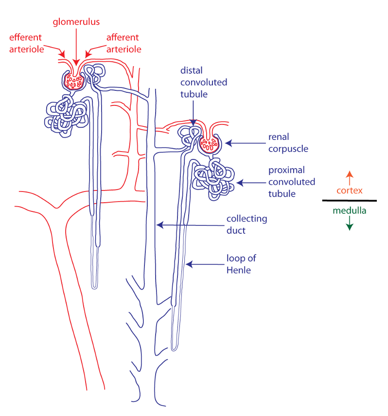
The first part of the nephron is the renal corpuscle, which consists of a tuft of the capillaries, the glomerulus (red), surrounded by Bowman’s capsule (blue). The rest of the nephron is called the renal tubule. The entire renal tubule is formed by a simple epithelium, and epithelial transport is important in the function of the renal tubule. The initial part of the renal tubule is the highly twisted proximal convoluted tubule (or proximal tubule), located in the cortex. This is followed by the loop of Henle, a straight segment that dives deep into the medulla, makes a hairpin turn, and then ascends back toward the cortex. The next segment, also in the cortex, is the smaller distal convoluted tubule (or distal tubule). Connecting tubules (not labeled) link the distal tubule to the collecting duct, a structure that is found in both the cortex and the medulla.
The nephrons use three basic processes in the homeostatic regulation of the extracellular fluid, with the result being the formation of urine that drains from the collecting ducts. These processes are depicted in the schematic nephron below, which has been untwisted and simplified to emphasize these processes.
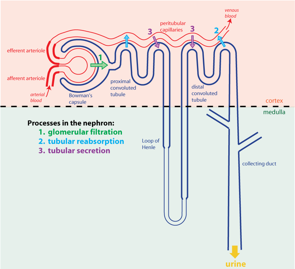
First, blood plasma is filtered through a nonspecific process of bulk flow known as glomerular filtration. This occurs in the renal corpuscle when the fluid and small molecules dissolved in plasma move from the inside of the glomerular capillaries into Bowman’s space, which is contained within Bowman’s capsule.
Next, the forming urine (or filtrate) flows through the renal tubule. In the renal tubule, water and substances that the body needs to keep will be transported out of the lumen of the tubule and back into the extracellular fluid; this process is called reabsorption. Certain substances that need to be eliminated from the body are transported from the extracellular fluid into the lumen of the tubule; this process is called secretion. Both reabsorption and secretion depend on epithelial transport. The movement of water depends upon osmosis.
Structures in the Renal Corpuscle
The renal corpuscles are where filtration occurs. Renal corpuscles are found exclusively in the cortex. The figure below shows a low-magnification view of the kidney cortex.
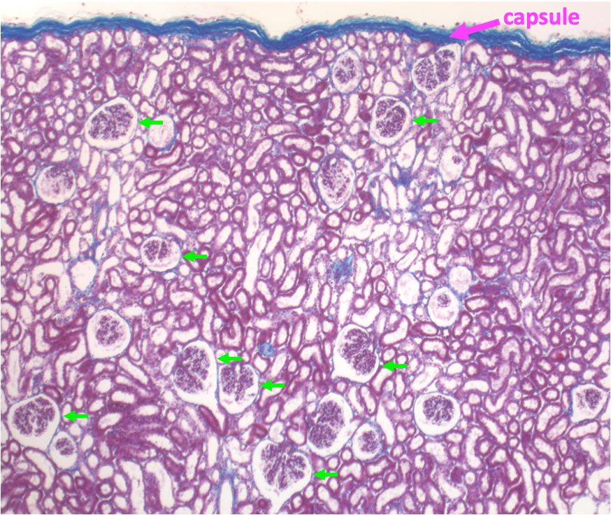
The renal corpuscles are the blobs that are enclosed within the circular spaces. A further indication that this section is from the cortex (besides the presence of corpuscles) is the connective tissue capsule seen at the top edge of the tissue section.
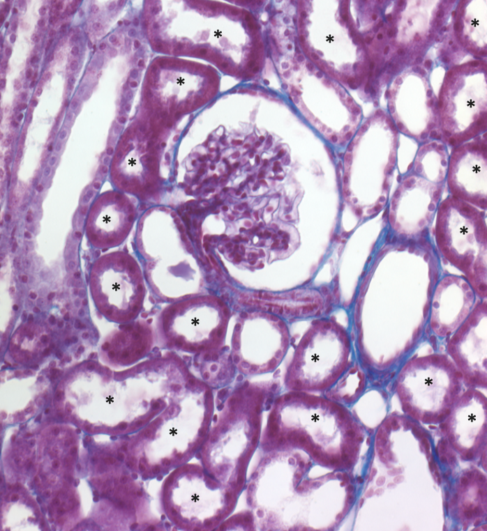
A higher magnification view of a renal corpuscle shows the glomerulus in the center, surrounded by Bowman’s capsule.
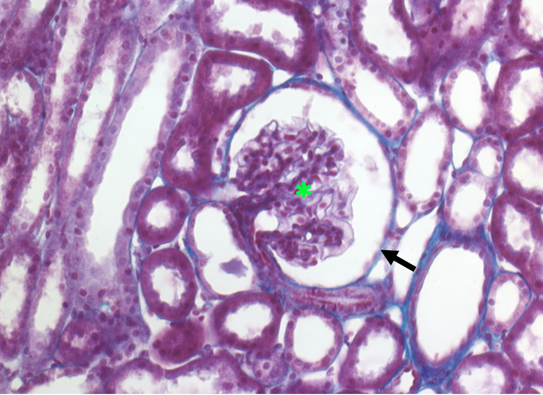
The outside of Bowman’s capsule, which encloses Bowman’s space, is formed by a simple squamous epithelium. In the tissue surrounding the renal corpuscle, the circles and ovals formed by simple cuboidal epithelium are sections through the renal tubule, mainly sections of proximal tubules and distal tubules (see later in the chapter).
Structures Forming the Filtration Membrane
The figure below focusses in on the part of the schematic nephron depicting the renal corpuscle.
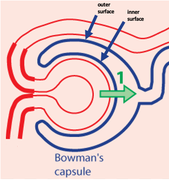
In this figure, the glomerulus is simplified so that it appears as a single capillary loop. Note that Bowman’s capsule consists of two surfaces. One surface is the simple squamous epithelium that we just saw in the previous figure. This surface forms the outside of Bowman’s capsule and encloses a space called Bowman’s space. The other surface of Bowman’s capsule is the inner surface (also called the visceral surface) that surrounds the glomerular capillaries. The inner surface of Bowman’s capsule is made up of a simple epithelium of podocytes, cells that wrap around the glomerular capillaries and form a part of the filtration membrane.
In the figure below, the podocytes are the pale lavender cells in the illustration of a renal corpuscle on the left. The podocytes completely cover the endothelial cells making up the glomerular capillaries.
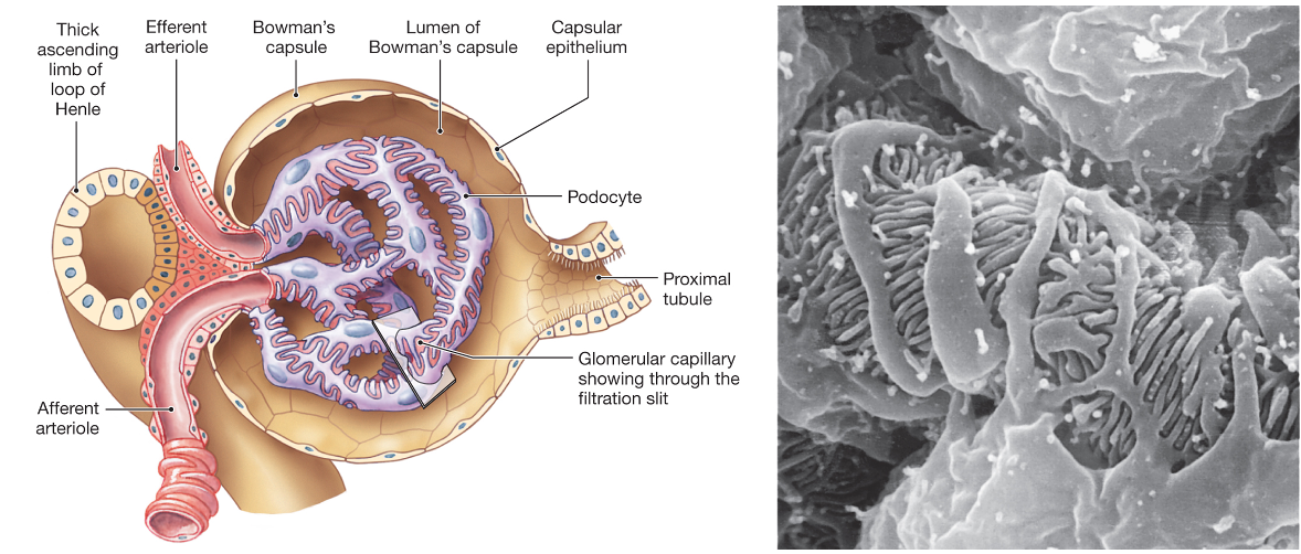
The figure on the right is a scanning electron micrograph of the outer surface of a glomerular capillary, showing podocytes. Podocytes have processes called arms that extend from their cell bodies. Along the arms are smaller processes called foot processes that interdigitate with other foot processes. (“Podo-” “-cyte” literally translates to “foot cell”.)
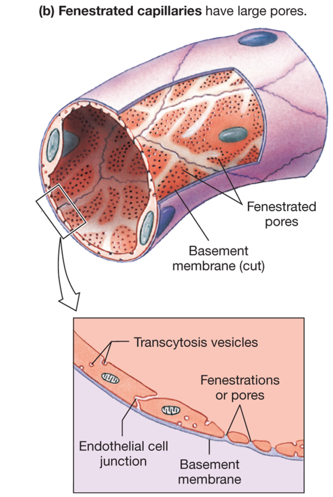
The endothelial cells that make up the glomerulus are specialized to favor filtration. The glomerular capillary endothelial cells have fenestrations, which are pores that pass entirely through the cell. The figure below illustrates the structure of a fenestrated capillary.
The figure below shows another scanning electron micrograph of a glomerulus. This micrograph was prepared using the technique of freeze-fracture replication, in which frozen tissue is fractured, coated with heavy metals, and imaged using scanning electron microscopy. The advantage of this technique is that it allows different surfaces of the tissue to be visualized.
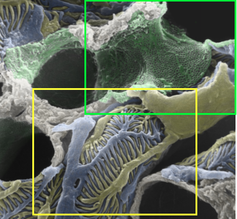
In this figure, the fracturing allows us to see both the inside and the outside of the glomerular capillaries. The above figure is pseudo-colored to help distinguish the inside of the capillary (green) from the outside covered by two podocytes (blue and yellow). The green and yellow boxes indicate the regions that are enlarged in the next figures.
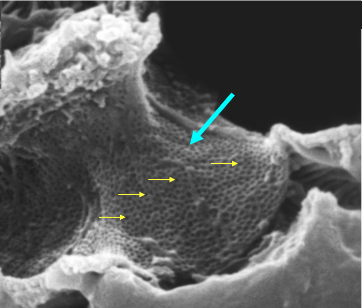
The figure below (not pseudo-colored) shows the enlarged inner surface of the capillary. The many small circles visible on the inner surface of the endothelial cell are fenestrations.
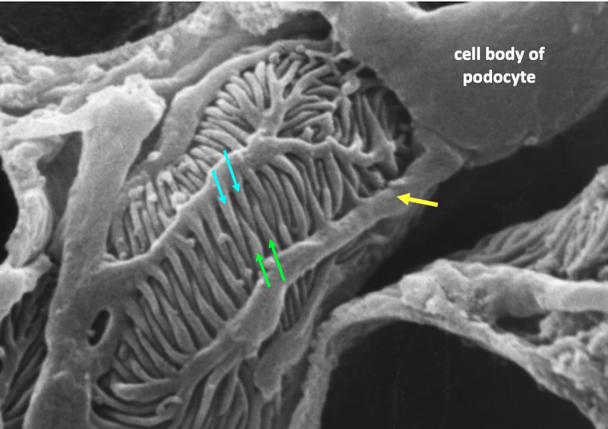
This figure shows the enlarged outer surface of the capillary, showing in detail a podocyte, its arms, and its foot processes.
The next figure shows how these two epithelia (the fenestrated endothelium of the capillary and the podocyte layer) combine to form the filtration membrane.
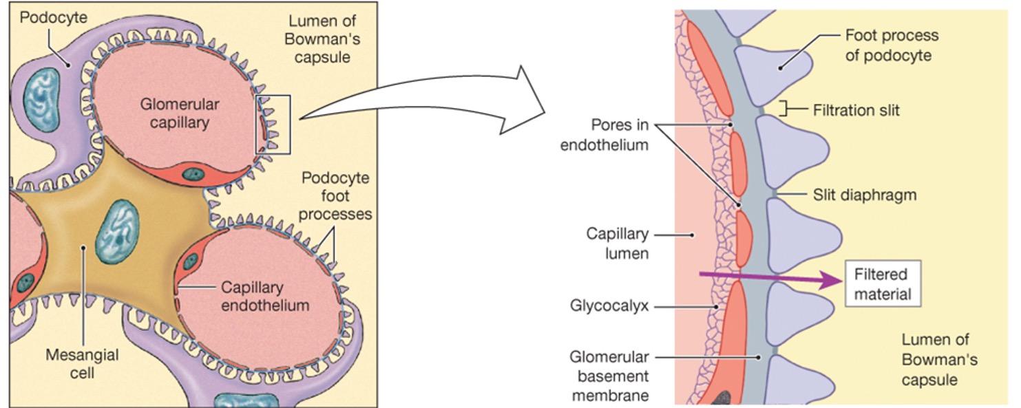
On the left is a cross section showing two capillary loops within the glomerulus. On the right is a highly magnified view of the filtration membrane. The endothelium and the epithelium created by the podocytes have between them a shared basement membrane called the glomerular basement membrane. The path of a filtered substance is first through a fenestration, then through the glomerular basement membrane, and finally through a filtration slit, which is the space between the podocyte foot processes. A complex of proteins called the slit diaphragm spans the filtration slit.
Structures in the Renal Tubule
After moving into Bowman’s space, the forming urine (or filtrate) flows into the renal tubule. The renal tubule is a hollow structure whose walls are formed by a simple epithelium. Through most of the renal tubule, this is a simple cuboidal epithelium, the exception being part of the loops of Henle. Different regions of the renal tubule express specific transport proteins and are key for certain processes. The functions of each part of the nephron are summarized in the figure below.
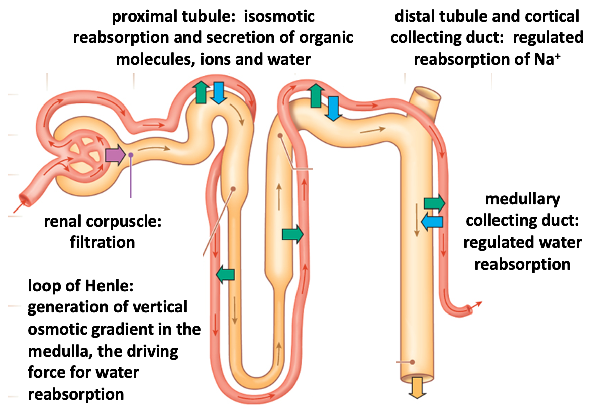
We will examine the histology of two parts of the renal tubule: the proximal tubule and the collecting duct.
Proximal Tubule
The figure below shows a section through the renal cortex, which contains renal corpuscles as well as proximal and distal tubules and the first part of the collecting duct. This figure captures Bowman’s space opening up into a proximal tubule.
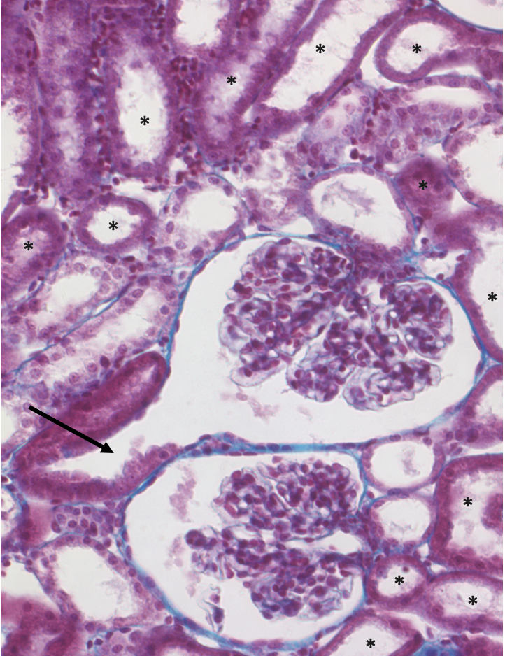
The proximal tubule is the major site for reabsorption and secretion in the renal tubule. Because these are energy-dependent processes that utilize ATP, the proximal tubule is full of mitochondria. In these two light micrographs, the mitochondria stain with the bright pink dye, and so the proximal tubules are the circles and ovals whose epithelium is more intensely stained. The cuboidal epithelium in the proximal tubule also has a fuzzy appearance due to the fact that the cell membranes are highly folded. The lighter stained tubules are distal tubules and potentially a few cortical collecting ducts.
The next figure is a transmission electron micrograph of the proximal tubule epithelium.
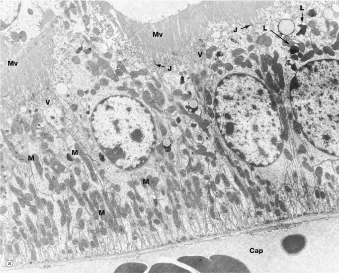
The electron micrograph shows that proximal tubule cells are filled with mitochondria, which appear as grey ovals in the cytoplasm of the cells. The apical membrane (at the top) is folded into microvilli, which increases the amount of surface area available for the expression of membrane transport proteins. The basolateral membrane (bottom) is also folded into larger membrane processes.
Collecting Duct
The collecting duct is involved in regulated reabsorption of sodium and water and is also involved in acid-base balance. We will examine the medullary collecting duct, which plays a key role in the regulated absorption of water.
To look at the histology of the collecting duct, we will take advantage of the structure of the rodent kidney, which, unlike a human kidney, has a single papilla. This means that a cross-section of the tissue will be organized into distinct regions of cortex, outer medulla (closest to the cortex), and inner medulla (closest to the papilla).
The inner part of the medulla contains only collecting ducts and thin segments of the loop of Henle. The thin segments of the loop of Henle are made up of simple squamous epithelium, while the epithelium of the collecting duct is simple cuboidal epithelium.
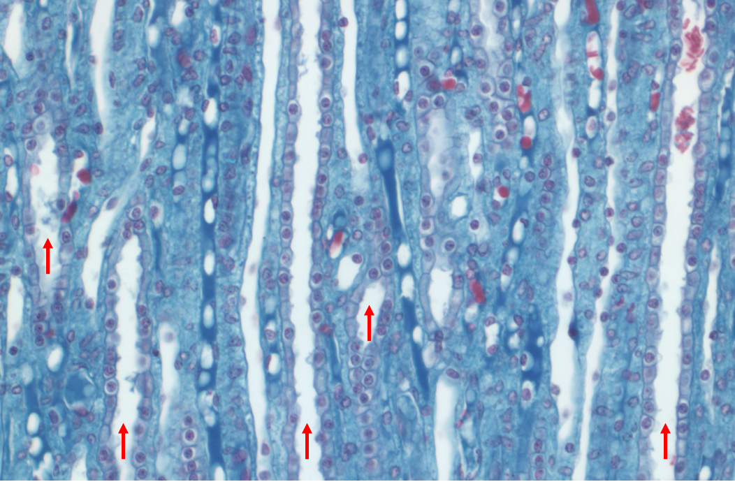
The high powered micrograph above shows several collecting ducts. A characteristic of the collecting duct is the crisp, clear outlines of the cells in the cuboidal epithelium.
Histology of the Collecting System and Bladder
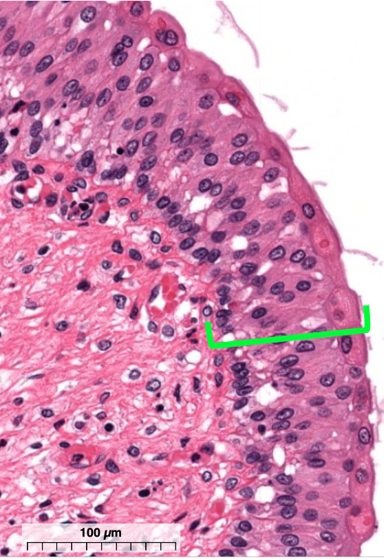
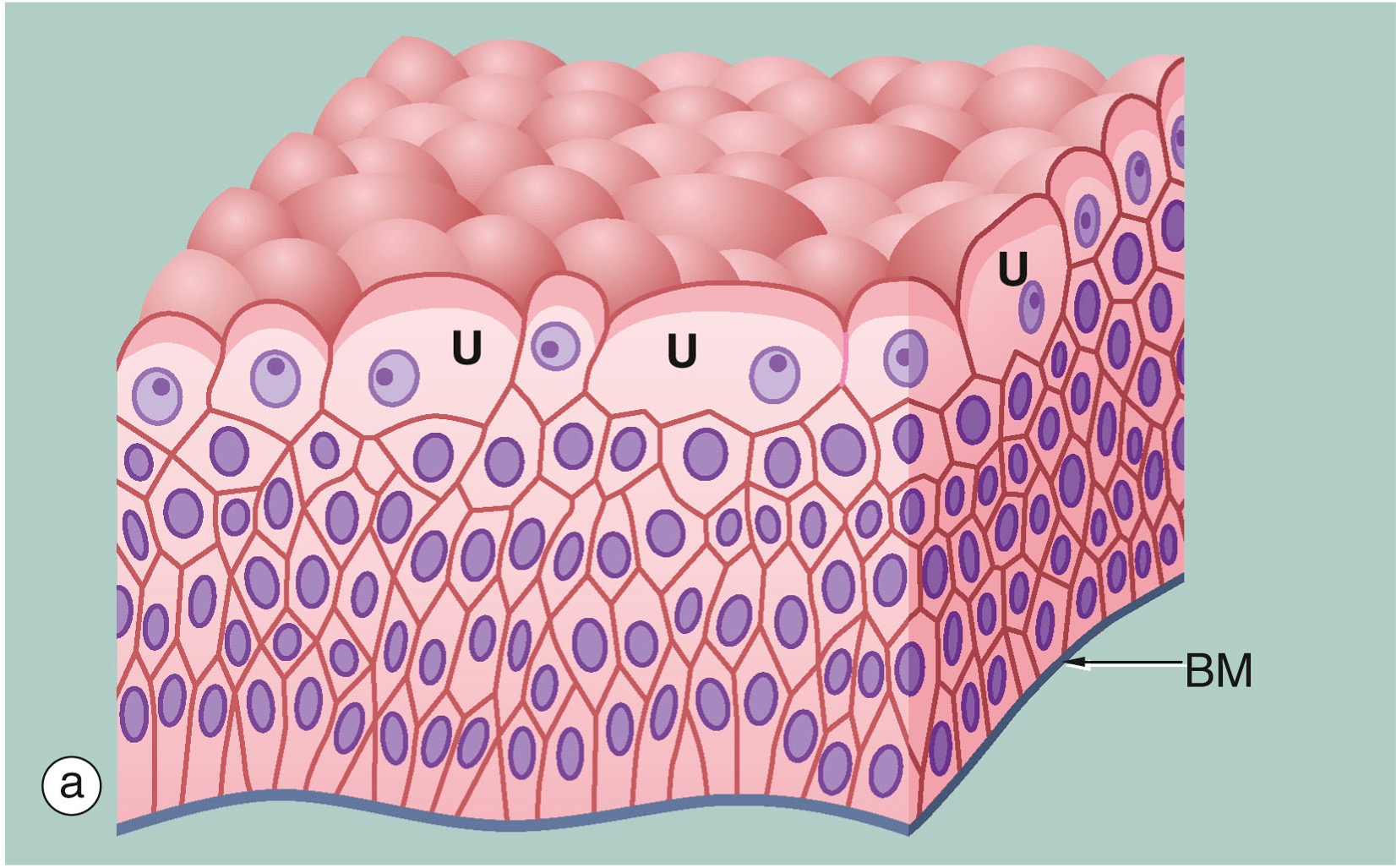
The collecting system of the kidney and the bladder are lined with a special type of stratified epithelium called uroepithelium or transitional epithelium. (Recall that a stratified epithelium is one that is made up of multiple layers of cells).
The uroepithelium possesses two key features:
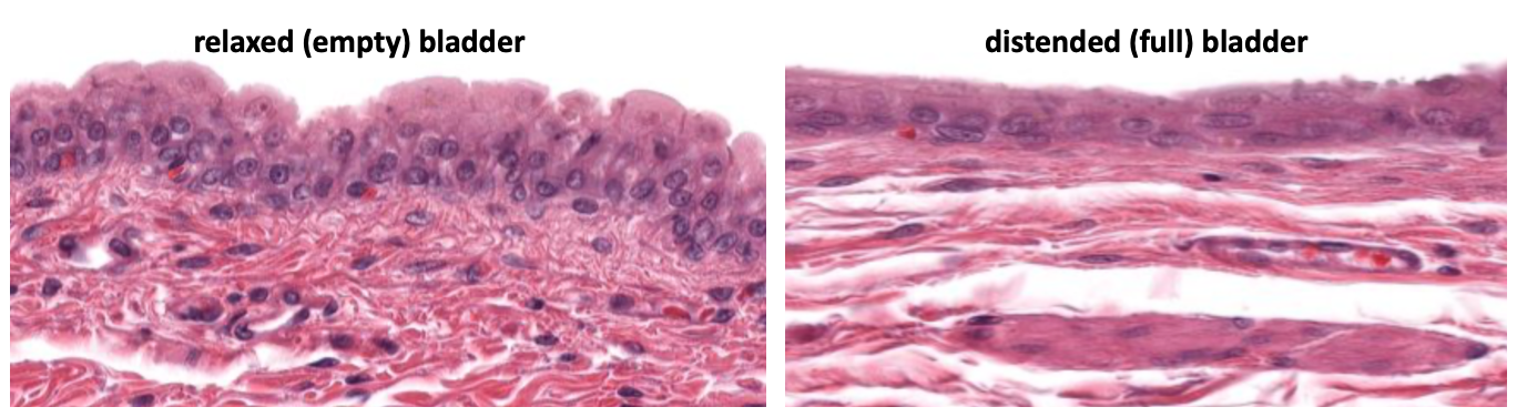
- It can adapt to the changing volume of the bladder and collecting structures. The uroepithelium stretches when these structures are full of urine and distended, and it folds up like an accordion when these structures have a small volume. There are specialized cells called umbrella cells (labeled with a U in the figure above) that are located at the apical surface of the uroepithelium that can change their size and shape as structures fill with urine.
- It is largely impermeant to water and toxins. Thus concentrated or dilute urine maintains its state while it is being stored in the bladder. Umbrella cells are important in the barrier function of the uroepithelium.
The figure below illustrates the transitional nature of the uroepithelium in the bladder, comparing its appearance in a relaxed and distended state.
The first example of uroepithelium that we will examine is from a calyx in a rodent kidney. At low magnification, one can see the papilla, containing sectioned collecting ducts, as it fits into the calyx.
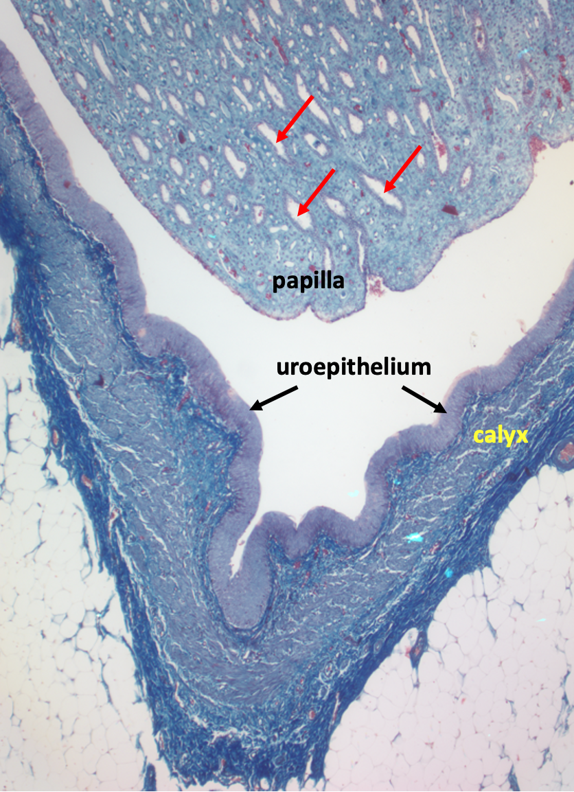
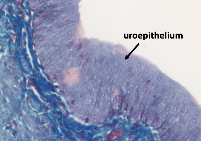
The calyx is lined with uroepithelium, which is shown at high magnification in the figure below.
The next figure shows a low magnification view of a section through the entire bladder.
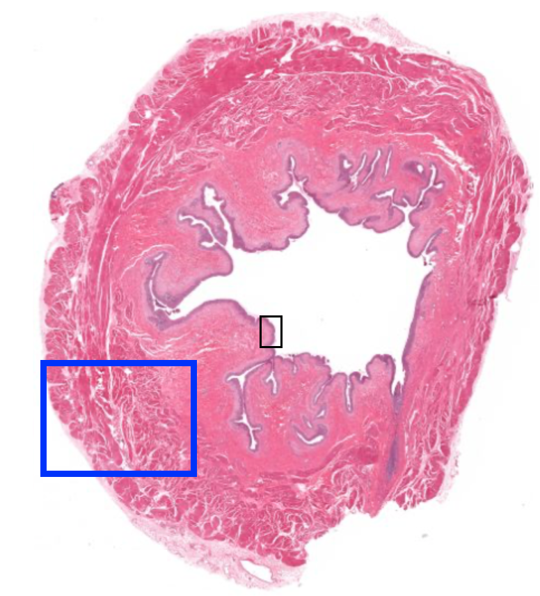
https://histologyguide.com/slideview/MHS-214-bladder/16-slide-1.html
The bladder wall is mostly made up of irregularly arranged smooth muscle which is known as the detrusor muscle.
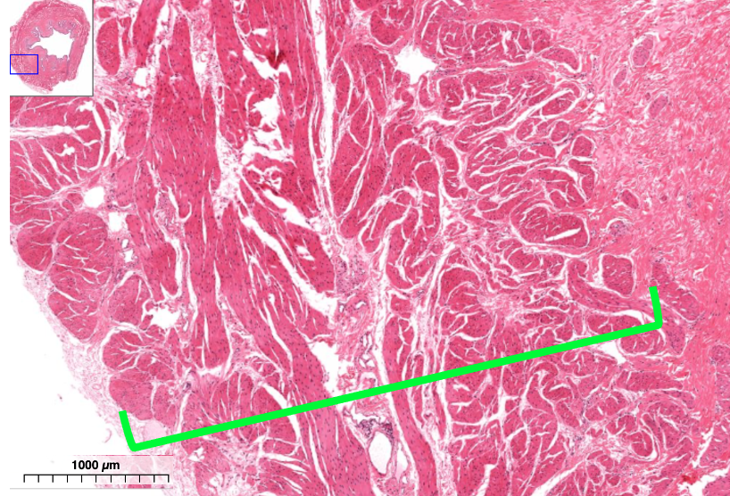
The bladder wall also contains some connective tissue. The inner surface of the bladder is lined with uroepithelium.