17 Skeletal Muscle Anatomy: Arm and Leg
We will be studying muscle anatomy using anatomical models of the arm and leg. The idea is to give you a flavor of how to study anatomy and to introduce you to the language used in naming skeletal muscles (Latin).
In gross anatomy, skeletal muscles have a fibrous appearance due to the presence of fascicles within the muscle.
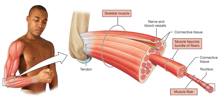
Fascicles are bundles of muscle fibers. Connective tissue wraps around individual muscle fibers, fascicles, and the outside of the muscle. Use the pattern of the fascicles to help distinguish the different muscles.
Muscles of the Arm
Muscles that move the arm at the shoulder
The muscles that move the arm at the shoulder by necessity have their origins on the trunk. These are pectoralis major, deltoid, and latissimus dorsi. The deltoid is an important muscle to know because intramuscular vaccination injections are usually administered into the deltoid.
These muscles are illustrated in the two drawings below.
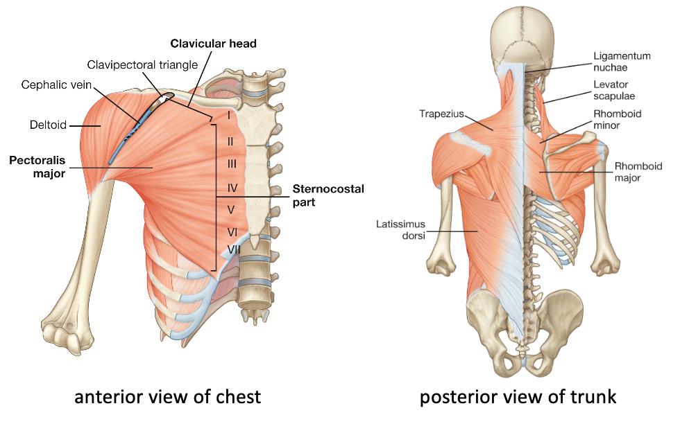
The arm model does not include pectoralis major and latissimus dorsi, so we will study the structure of these muscles using a required video from the Acland’s Video Atlas of Anatomy. The link is below.
1.1.9 Latissimus dorsi, pectoralis major, and deltoid muscles
This figure below provides two key views of these muscles from screenshots in the video.
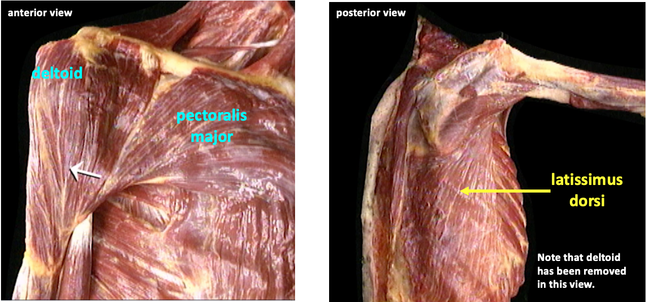
The actions of pectorals major are medial rotation and adduction of the arm at the shoulder. Remember that with adduction you are “adding” the limb back to the body. Pectoralis major also lifts the arm forward in the anterior-posterior plane; this action is flexion of the arm at the shoulder.
Latissimus dorsi also causes medial rotation and adduction of the arm at the shoulder. (That latissimus dorsi causes medial rotation is not very intuitive without knowing that it attaches toward the front of the humerus and thus can turn the arm medially. Note that in the Acland video he refers to medial rotation as “internal rotation”). By virtue of its location on the back, latissimus dorsi causes extension of the arm at the shoulder–essentially pulling the arm back after flexion.
The deltoid muscle has many actions because although it is one muscle, it has anterior and posterior groups of fibers that can be activated independently. Thus, deltoid causes both medial and lateral rotation, and both flexion and extension of the arm at the shoulder. If all the fibers of deltoid are active together, it causes abduction of the arm at the shoulder (lifting the arm out from the side in the frontal plane).
Muscles that move the forearm
Muscles located in the upper arm are important for flexion and extension of the forearm at the elbow. The Latin root meaning “upper arm” is “brachi-“, which forms a part of the name for most of these muscles.
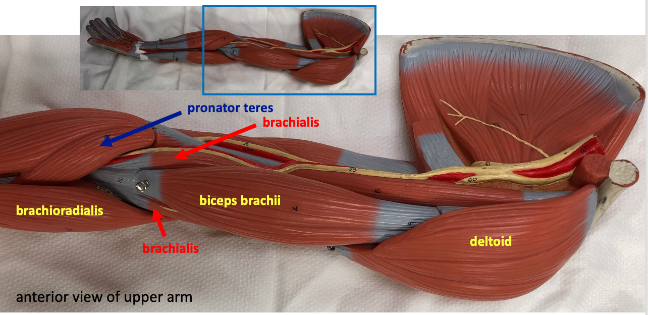
In the anterior view of the arm model above, it is possible to see the deltoid muscle, and the three flexors of the elbow: biceps brachii, brachialis, and brachioradialis. The term “biceps” means having two “heads” or two origins. This isn’t immediately obvious because where the two origins of biceps brachii divide is mostly hidden by deltoid. Brachialis is a deep muscle, and only visible on either side of biceps brachii, the superficial muscle. Brachioradialis originates lower on the humerus and inserts on the radius; hence its name tells you the origin and the insertion of this muscle. The action of these muscles is to flex the arm at the elbow.
Another muscle visible in this view is pronator teres. This muscle is named according to its action, which is pronation of the forearm (medial rotation of the forearm, bringing the thumb around medially). The diagonal orientation of pronator teres provides a hint about its action.
The muscle that causes extension of the forearm at the elbow is triceps brachii, which is visible on the posterior side of the upper arm.
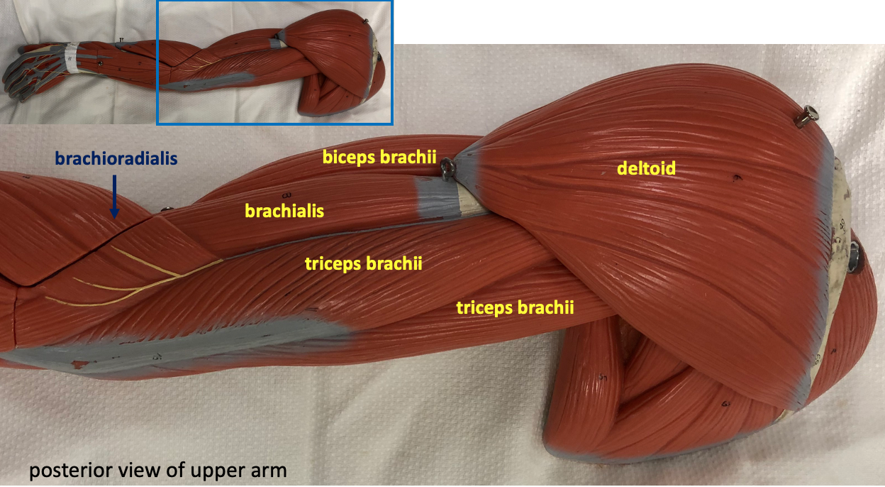
Triceps brachii has three origins, although only two are visible in this view. There is also a good view of brachialis and brachioradialis from the posterior side of the upper arm.
Muscles that move the wrist
Muscles located on the forearm (lower arm) are involved in moving the fingers and the wrist. To identify these muscles, you need to know that flexion of the wrist involves bending the wrist so that the fingers move towards the forearm; extension of the wrist is the opposite movement. To achieve flexion, the muscle tendons need to insert on the anterior side of the wrist, thus the muscles on the anterior forearm are flexors.

We are just going to learn to identify the three superficial muscles on both the anterior and posterior surface of the forearm. Conveniently, most of these muscles are named according to their actions. In the anterior view above, the lateral muscle, positioned over the radius, is called flexor carpi radialis. “Carpi” is Latin for “wrist”. The medial muscle, positioned over the ulna, is called flexor carpi ulnaris.
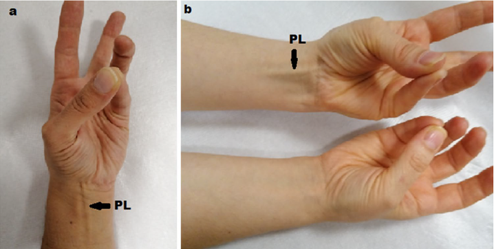
The central flexor muscle on the surface of the forearm is called palmaris longus. This muscle is quite frequently absent, without having any effect on grip strength or function. The figure below shows a simple test that accentuates the palmaris longus tendon, and can be used to show whether the muscle is present or absent.
Muscles whose tendons insert onto the posterior side of the wrist cause extension of the wrist.
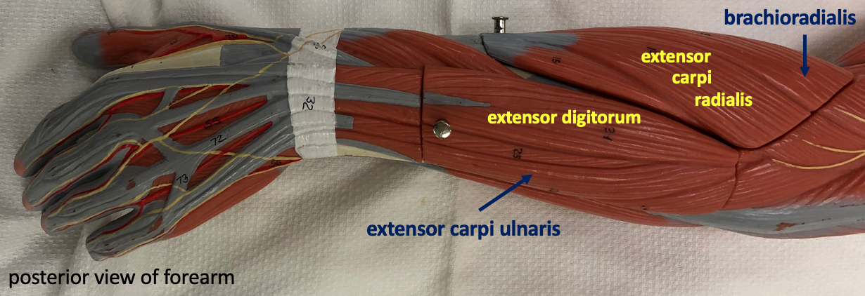
Similar to the names of their counterparts on the anterior side, the lateral and medial muscles are extensor carpi radialis (lateral) and extensor carpi medialis (medial). Extensor carpi radialis (to be precise, the muscle labeled is extensor carpi radialis longus; extensor carpi radialis brevis is mostly not visible) has its origin quite close to the origin of brachioradialis, and the two muscles run in parallel. The muscle in the middle is extensor digitorum. Note how its tendons connect to the bones in the fingers. In addition to causing extension of the wrist, extensor digitorum also causes extension of the digits.
Muscles of the Leg
Muscles that move the thigh
The muscles we are learning about that move the thigh have their origins on bones of the pelvis. The tensor fasciae latae is a muscle on the anterior-lateral side of the thigh that causes abduction of the thigh. This muscle also tenses fascia lata (as indicated in its name). The fascia lata is a wide sheet of connective tissue that covers the muscles of the thigh. The figure below illustrates the fascia lata.
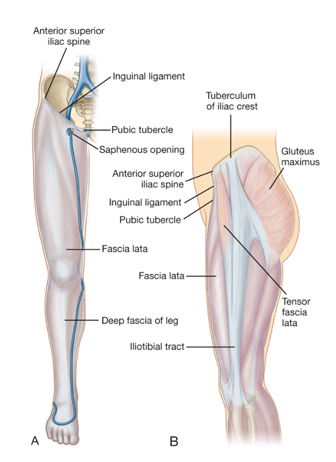
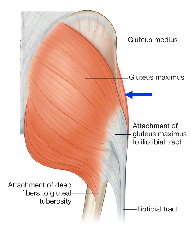
On the lateral side of the leg, the fascia lata thickens to form a structure known as the iliotibial tract, or iliotibial band. The iliotibial tract is located in the middle of the lateral side of the leg, just where the seam of one’s pant leg would be. The iliotibial tract provides an insertion for tensor fasciae latae, but also for gluteus maximus, as shown in the figure above.
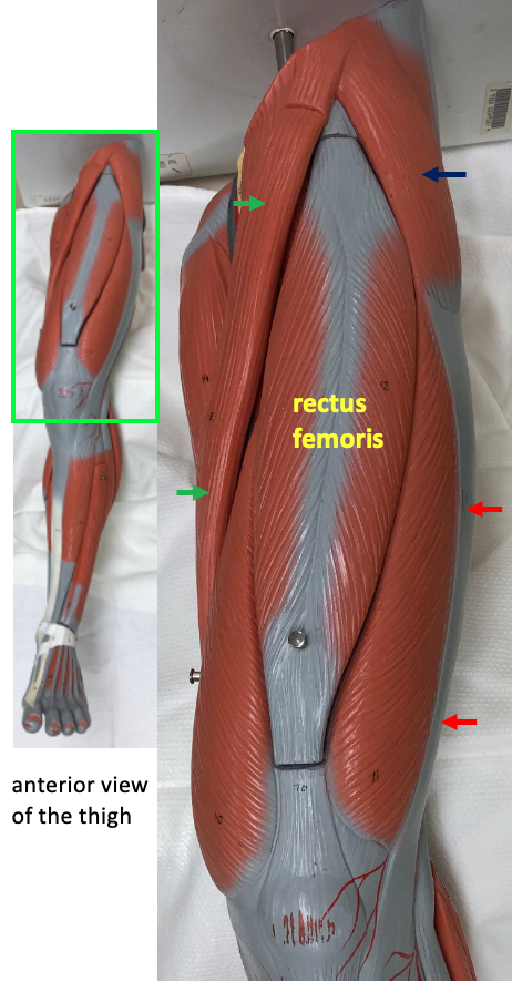
On the anterior side of the thigh are two muscles that flex the hip at the thigh: rectus femoris and sartorius. “Rectus” means “straight” (think of the word direct). Rectus femoris actually crosses both the hip joint and the knee joint, and so is involved in extension of the lower leg as well as flexion at the hip. Sartorius is a thin, strap-like muscle that crosses the thigh diagonally and also crosses both the hip joint and the knee joint. In addition to weak flexion of the hip, sartorius also causes weak lateral rotation at the hip, and weak flexion of the knee.
The dominant muscle on the surface of the buttocks is gluteus maximus. This muscle is a very powerful extensor of the thigh. You use gluteus maximus when you are climbing stairs or lowering your body into a chair. As shown in the illustration below, one of the insertions of gluteus maximus is the iliotibial tract.
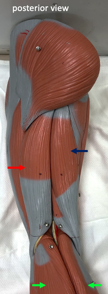
The other muscles that cause extension of the thigh at the hip are the muscles of the hamstring group: biceps femoris, semitendinosus, and semimembranosus. The name “hamstring” is related to the long, cord-like tendons for these muscles that are present on the posterior side of the knee.
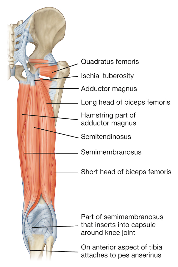
All of the muscles in the hamstring group have origins on the ischial tuberosity and insertions on the bones of the lower leg. The two medial muscles are semimembranosus, which is deep (and not very visible on the model), and semitendinosus, which is on the surface. The lateral muscle, biceps femoris is so called because it has two origins: one on the ischial tuberosity and one on the femur. The hamstring muscles cross both the hip and the knee joint, and so they cause flexion of the lower leg at the knee, as well as causing extension at the hip.
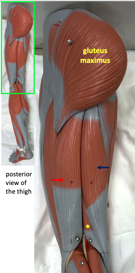
The extensors of the thigh are visible in the posterior view of the thigh shown below. Note that the drawing in the figure above depicts a right leg, while the model shown below depicts the left leg.
The muscles found in the groin region act as adductors of the thigh: they bring the leg toward the midline of the body. The three major adductors are adductor magnus, adductor longus, and gracilis.
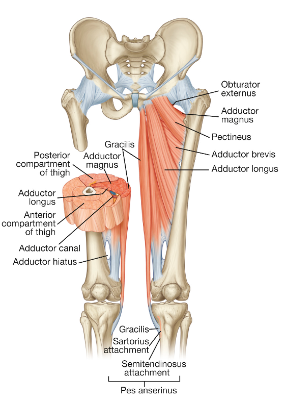
The adductor muscles are best seen in a medial view of the thigh.
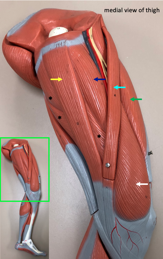
Although adductor magnus is a large muscle, it is mostly hidden in this model by gracilis, a strap-like muscle that extends from the pubis to the tibia. Adductor longus is visible at the top of the thigh in the triangular space between sartorius and gracilis.
Muscles that move the lower leg
There are four muscles in what is called the quadriceps group (quadriceps meaning “four heads” for the four origins of the four muscles). The quadriceps muscles are responsible for extension of the lower leg at the knee. These muscles are depicted in the drawing below.
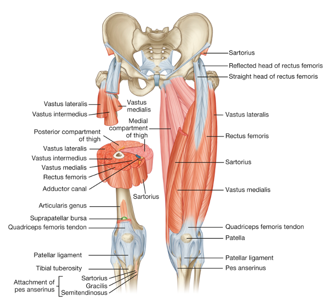
Three of the quadriceps muscles are visible on the surface of the thigh: rectus femoris, vastus medialis, and vastus lateralis. Vastus intermedius lies between vastus medialis and vastus lateralis, and deep to rectus femoris. Its location can be seen in the sectional view of the muscles in the drawing above. The quadriceps muscles all attach to the patellar tendon, which inserts on the tibia at the tibial tuberosity.
Below is the anterior view of the thigh, but with the focus now on the quadriceps muscles that are visible.
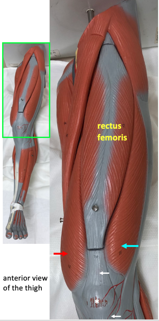
The stretch reflex (see next chapter) is often tested at the knee. A doctor will use a reflex hammer to hit the patellar tendon in the spot just below the patella. This causes a sharp stretch in the muscles of the quadriceps group, and the reflex response activates contraction in these muscles to cause extension of the lower leg.
The hamstring muscles (semimembranosus, semitendinosus, and biceps femoris) are responsible for flexion of the lower leg at the knee. The photo below shows those muscles on the posterior thigh, but also includes the top of gastrocnemius, a muscle found on the surface of the calf. Because of its origin on the femur, gastrocnemius causes weak flexion of the knee, but its main action is plantar flexion (see below).
Muscles that move the foot
The actions at the ankle joint have specific names. For instance, to describe the action that is pointing of the toes, rather than saying “extension of the foot”, we say “plantar flexion“. The opposite movement to plantar flexion is dorsiflexion, which involves bending the ankle to bring the toes toward the shin.
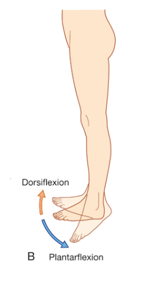
The muscles on the posterior part of the calf are responsible for causing plantar flexion.
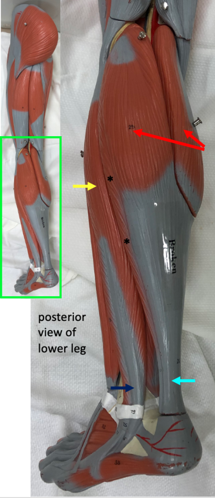
One of these muscles is gastrocnemius, a large muscle with two prominent oval heads, each of which connects to a broad flat tendon below. The soleus muscle is a large flat muscle that is not very visible because it lies deep to gastrocnemius. Both gastrocnemius and soleus insert onto the calcaneus via the Achilles tendon (also called the calcaneal tendon). The muscles that perform plantar flexion are large and powerful because plantar flexion is the propulsive movement during walking and running. The muscles of plantar flexion push against the ground to lift the whole body; by contrast, the muscles that perform dorsiflexion only need to lift the foot.
The muscle on the lateral surface of the fibula, fibularis longus (also called peroneus longus) performs plantar flexion because its tendon attaches on the bottom of the first metatarsal after curving around behind the lateral malleolus. Fibularis longus also performs eversion of the foot. This is the action that turns the sole of the foot outward. Eversion, and the corresponding opposite movement, inversion, are important for keeping the body stable and upright on an angled surface.
The anterior view of the lower leg shows the muscles that cause dorsiflexion. As mentioned above, dorsiflexion only requires the lifting of the foot, so these muscles are much smaller than the powerful muscles on the back of the calf that perform plantar flexion.
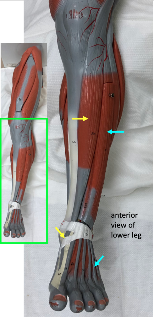
Tibialis anterior has its origin on the lateral front surface of the tibia. Its tendon inserts on the first metatarsal (the instep of the foot). In addition to dorsiflexion, tibialis anterior also causes inversion of the foot, which is the turning of the sole of the foot inward (medially). Assisting tibialis anterior with dorsiflexion is the muscle extensor digitorum longus. The tendons of this muscle fan out to the four small toes. As per its name, in addition to dorsiflexion, extensor digitorum longus causes extension of the toes.
