12 Bones of the Lower Limb

Bones of the Lower Limb

For reference, the figure below shows how all the bones of the lower limb fit together.

photo and illustration of the bones of the leg
Anterior view of the arrangement of bones in the pelvis and leg. Figure 6.1 in Human Anatomy, Color Atlas and Textbook by J.A. Gosling et al., 6th edition (2017).

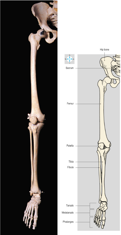
Just as the arm is attached to the trunk via the shoulder girdle, the leg is attached to the trunk via the pelvic girdle.  There are three bones that make up the pelvis:  two hip bones anteriorly, and the sacrum posteriorly.

anterior view of pelvis
Anterior view of a plastic model of the pelvis. The bright green arrow indicates the pubic symphysis.

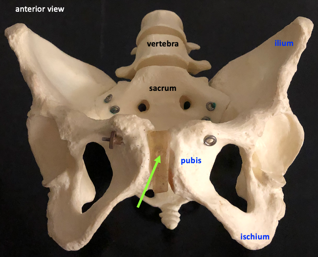
The hip bone (or os coxae–a term I don’t expect you to learn for this class) is actually the fusion of three bones:  the ilium, the ischium, and the pubis.  The anterior joint between the two pelvic bones consists of a fibrocartilage pad and is called the pubic symphysis.  This joint is only slightly mobile.

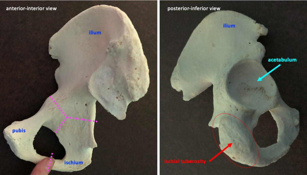
The figure below shows two views of the hip bone, and illustrates where the ilium, ischium, and pubis come together.  The three bones fuse in the acetabulum, the hip socket where the femur articulates with the pelvis.  On the inferior surface of the ischium is the ischial tuberosity, a rough area that is the origin (proximal attachment point) for the hamstring muscles (biceps femoris, semimembranosus, and semitendinosus).

interior view (left) and posterior-inferior view (right) of the hip bone, with lines to show the divisions between the three bones
Interior (left) and posterior-inferior (right) views of the hip bone. The dotted pink lines in the figure on the right indicate the divisions between the three bones.  The red oval in the figure on the right contains the ischial tuberosity.

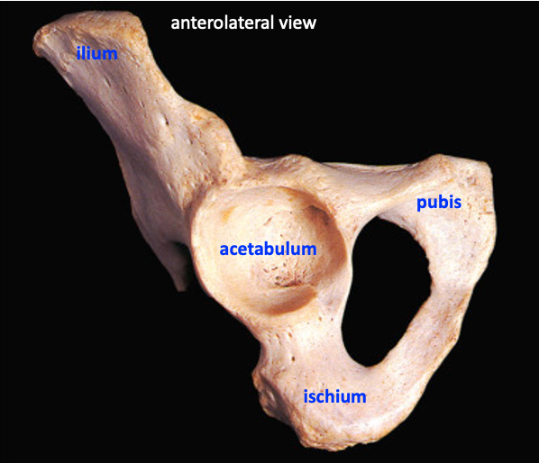
The figure below shows an anterolateral view of the hip bone with the acetabulum at center.

anterolateral view of hip bone
eral view of the hip bone showing the acetabulum. Adapted from Figure 6.26 in Human Anatomy, Color Atlas and Textbook by J.A. Gosling et al., 6th edition (2017).

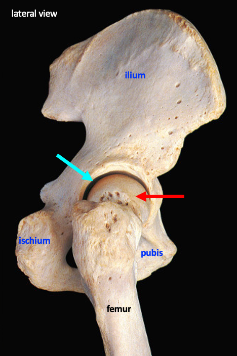
The next figure illustrates how the head of the femur articulates at the acetabulum to form the hip joint.

lateral view showing hip joint
A lateral view of the femur and hip bone forming the hip joint. The hip joint is a ball-and-socket joint where the head of the femur(red arrow) is the ball and the acetabulum (cyan arrow) is the socket.  Adapted from Figure 6.31 in Human Anatomy, Color Atlas and Textbook by J.A. Gosling et al., 6th edition (2017).

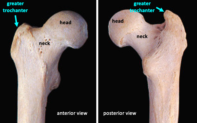
The femur is the thigh bone.  On its proximal end it has a round head that attaches to the rest of the bone by a skinnier neck.    The head articulates with the pelvis in the hip joint (see above).

anterior (left) and posterior (right) views of the femur
Anterior and posterior views of the femur. The proximal features of the femur are the head (red arrows), the neck (green arrows) and the greater trochanter (cyan arrows). At the distal end there is a large smooth area where the femur articulates with the patella and the tibia (asterisk in anterior view). In the posterior view, the medial condyle is indicated with a pink arrow and the lateral condyle is indicated with a yellow arrow. Adapted from Figure 6.16 (left) and 6.15 (right) in Human Anatomy, Color Atlas and Textbook by J.A. Gosling et al., 6th edition (2017).

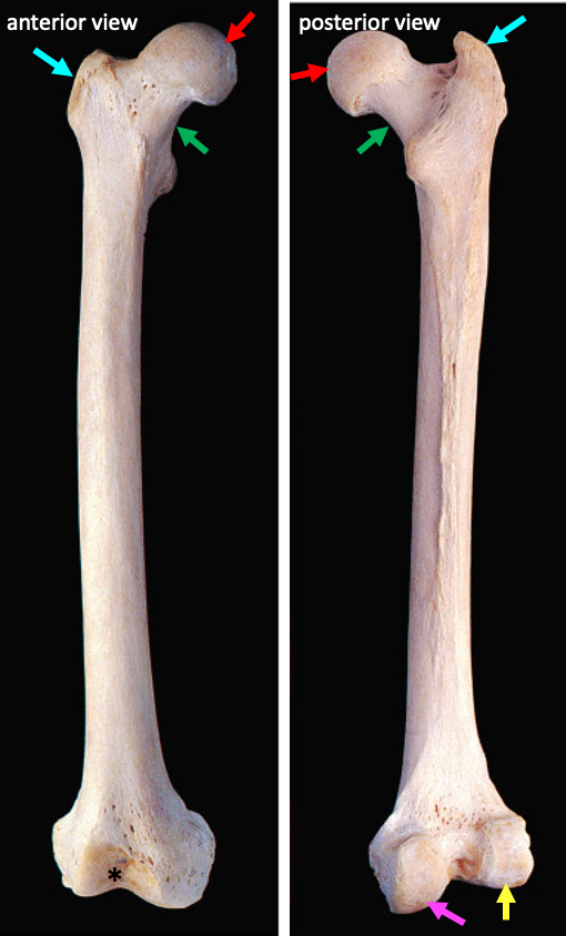
On the lateral side of the proximal femur is a large prominence called the greater trochanter.  The greater trochanter is an attachment site for muscles that move and stabilize the hip.

anterior (left) and posterior (right) view of the femur
Structures at the proximal end of the femur. Adapted from Figure 6.16 (left) and 6.15 (right) in Human Anatomy, Color Atlas and Textbook by J.A. Gosling et al., 6th edition (2017).

The distal end of the femur has a large smooth condylar surface where it articulates with the tibia and patella to form the knee joint.

bones of the knee joint (left) and articular surfaces of the femur (top) and tibia (bottom) on the right side
The knee joint. The left side is an anterior view showing the articulation of the femur, patella, and tibia. The right hand figure shows the articular surfaces of the femur and the tibia. Each bone has a lateral and medial condyle. Adapted from Figure 6.1 (left) and Figure 6.73 in Human Anatomy, Color Atlas and Textbook by J.A. Gosling et al., 6th edition (2017).

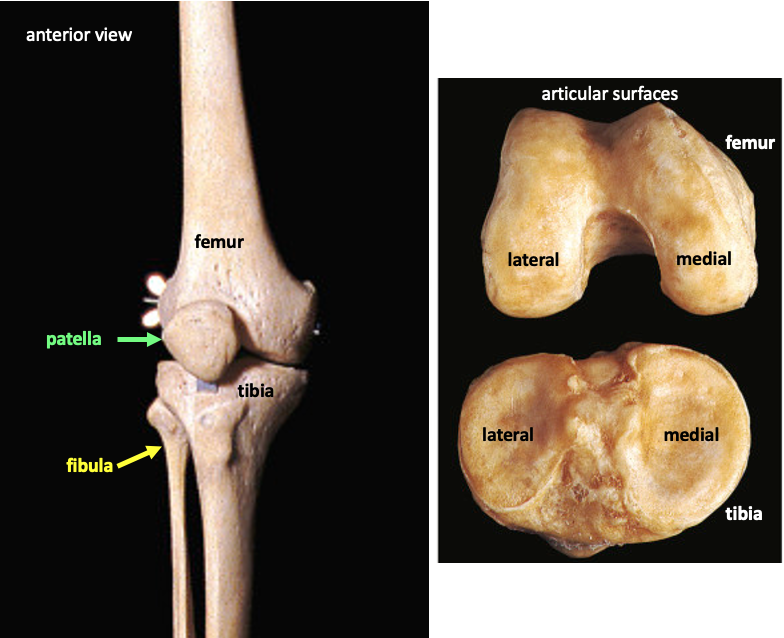
The condylar surface divides to form a medial condyle and a lateral condyle., which can be seen most easily in a posterior or inferior view of the bone.

posterior view of full femur (left) and enlarged version (right) showing medial and lateral condyles
Medial and lateral condyles at the distal end of the femur. Adapted from Figure 6.15 (right) in Human Anatomy, Color Atlas and Textbook by J.A. Gosling et al., 6th edition (2017).

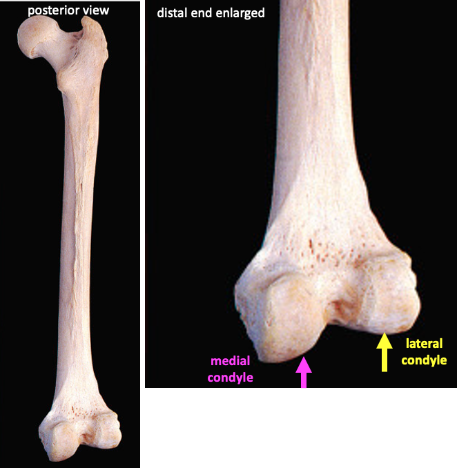
The medial and lateral condyles have a different appearance, but the way to confidently identify them is to look at the whole bone and note the medial side by located the head of the femur.

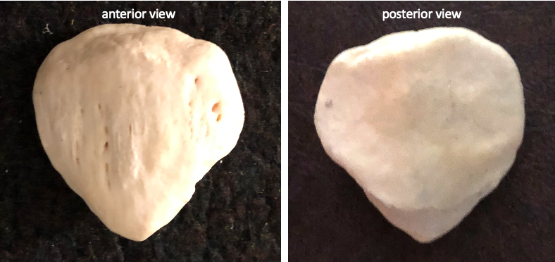
The patella (kneecap) is a sesamoid bone, meaning it is embedded in a tendon (the patellar tendon).

anterior (left) and posterior (right) views of the patella
The patella.  The posterior surface is smooth where it articulates with the femur.

The tibia (shinbone) is the large bone in the lower leg that supports the weight of the body and forms a joint with the femur at the knee.

bones of the lower leg and foot
Anterior view of the tibia (T) and the fibula (F) articulated with the bones of the foot. The yellow arrow indicates the medial malleolus on the tibia and the green arrowindicates the lateral malleolus on the fibula. Adapted from Figure 6.61 in Human Anatomy, Color Atlas and Textbook by J.A. Gosling et al., 6th edition (2017).

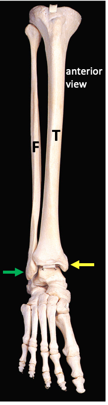
As with the femur, the condylar surface on the superior side of the tibia is divided into a medial condyle and a lateral condyle.  The way to confidently discriminate between medial and lateral condyles is to look to the distal end of the bone and identify the medial malleolus.  The medial malleolus forms the outer part of the ankle joint on the medial side.  This is the hard prominence you can feel under your skin.

anterior view of full tibia (left) and enlarged anterior-superior view showing medial and lateral condyles (right)
The medial and lateral condyles are on the superior surface of the tibia. The pink arrow indicates the lateral condyle; the cyan arrow indicates the medial condyle. The yellow arrow in the figure on the left indicates the medial malleolus. The asterisk indicates the tibial tuberosity.

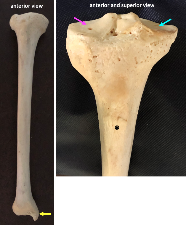
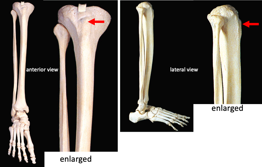
A distinguishing feature on the anterior surface of the tibia is the tibial tuberosity.  This is a rough prominence near the proximal end of the bone that is the attachment point for the patellar tendon, which links to the quadriceps muscles (vastus lateralis, vastus medialis, rectus femoris, and vastus intermedius).

anterior (left) and lateral (right) views of the lower leg, with enlarged insets to show the tibial tuberosity
Anterior (left) and lateral (right) views of the bones of the lower leg and foot. The tibial tuberosity is indicated by the red arrow. Adapted from Figure 6.61 (left) and Figure 6.65 (right) in Human Anatomy, Color Atlas and Textbook by J.A. Gosling et al., 6th edition (2017).

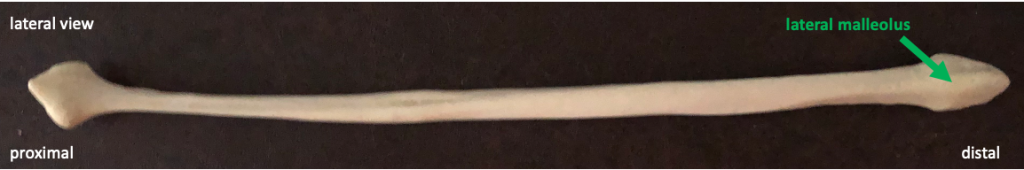
The fibula is the small lateral bone in the lower leg.  At its distal end is the lateral malleolus, which forms the outer part of the ankle joint on the lateral side.  Similar to the medial malleolus, it is easy to feel the lateral malleolus just under the skin at the ankle.

lateral view of the fibula in a horizontal orientation with proximal to the left
The fibula.
lateral view of the bones of the lower leg and foot
Lateral view of the bones of the lower leg and foot. The lateral malleolus of the fibula is indicated by the green arrow. The calcaneus is indicated by the blue arrow. Adapted from Figure 6.65 in Human Anatomy, Color Atlas and Textbook by J.A. Gosling et al., 6th edition (2017).

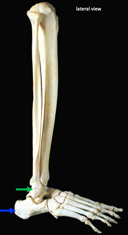
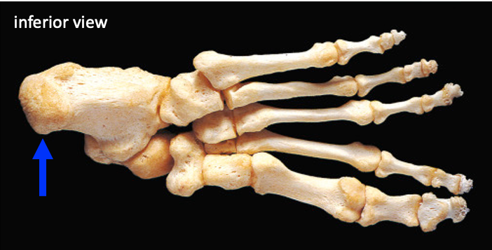
The calcaneus (heel bone) is the attachment for the Achilles tendon (the insertion for the gastrocnemius and soleus muscles).  The calcaneus is one of the tarsal bones.

inferior view of the bones of the foot, with the calcaneus (heel) on the left
The calcaneus (blue arrow) is the heel bone. Adapted from Figure 6.50 in Human Anatomy, Color Atlas and Textbook by J.A. Gosling et al., 6th edition (2017).

Optional Videos

Below are links to videos in the Acland’s Video Atlas of Anatomy.  They are  helpful in allowing you to get a sense of the bones in three dimensions.  They also will introduce you to the movements around the joints, which we will be studying in our class about muscle anatomy.  The first video listed below is the most useful since we are studying the anatomy of the knee joint.

2.2.1 Bony features of the knee joint

2.1.2 The hip bone

2.1.3 The femur

2.3.2 Bones and ligaments of the ankle joint


License

Human Physiology in Health and Disease (PBIO 375) Copyright © by Anna Melby. All Rights Reserved.

Share This Book