
14 Anatomy of the Knee Joint
Types of Joints
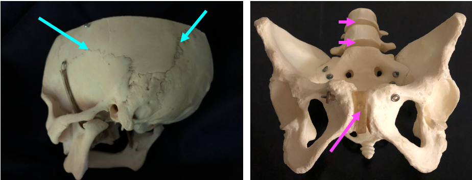
Joints (also called articulations) are the places where two skeletal element meet. The typical joints that we think of are highly movable joints known as synovial joints. But there are also immovable joints such as the sutures in the skull, in which two bones knit together by fibrous tissue that is eventually replaced by bone. As well, there are slightly movable joints such as the pubic symphysis or the joints between the vertebrae in the spine.
Synovial Joints
A synovial joint has a fluid filled space, the synovial cavity which is created because the area where the two bones meet is enclosed in a fibrous capsule. The inner surface of the joint capsule is lined by a membrane called the synovium. The synovium produces synovial fluid which fills the space of the synovial cavity.
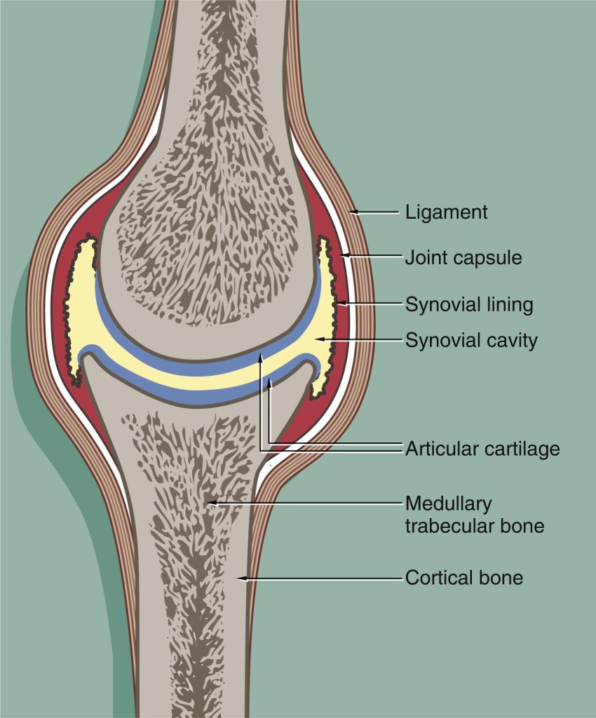
The ends of the bone that meet within the synovial cavity are covered by a layer of hyaline cartilage called the articular cartilage. In the dried bones of the skeleton that we examined last week, the very smooth surfaces of the condyles on the heads of the femur or humerus are places that in the living bone were covered with articular cartilage. The smooth articular cartilage combined with the lubrication of the synovial fluid provide a low-friction surface for movement between the bones in a joint.
Blood vessels do not penetrate into cartilage, and it has a limited ability for repair. This is particularly true for articular cartilage. Damage to articular cartilage occurs in arthritis.
Joints are stabilized by ligaments. A ligament is a piece of fibrous connective tissue that links bone to bone, while a tendon is a piece of fibrous connective tissue that links muscle to bone.
Knee Joint Anatomy
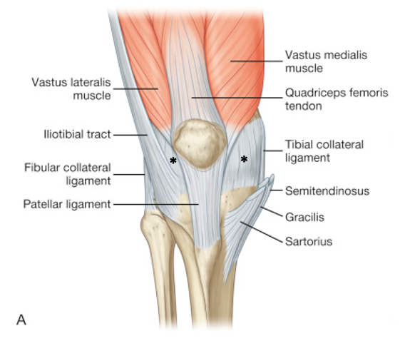
The figure below shows an anterior view of the knee joint where the fibrous capsule is intact.
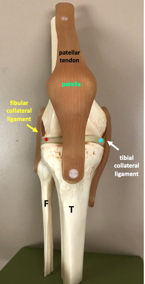
We will be studying a model of the knee joint in which the fibrous capsule is dissected away in order to reveal the inner structures of the knee joint.
The table below groups structures of the knee joint according to whether they are bone, cartilage, or ligaments.
| Bones | Notes | |
| femur |
large bone of the thigh | |
| patella |
the patella is contained within a large band of fibous connective tissue called the patellar tendon (or patellar ligament) | |
| tibia |
larger bone of lower leg that articulates with femur | |
| fibula |
use the fibula to know which is the lateral side | |
| Cartilage Structures |
||
| articular cartilage | articular cartilage is represented by the glossy surface on the femur, tibia, and patella | |
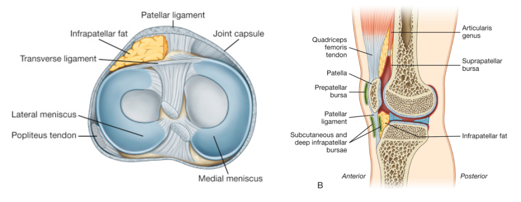
| medial meniscus | the meniscus is a wedge of fibrocartilage that helps to cushion the knee | |
| lateral meniscus | ||
| Tendons and Ligaments | ||
| patellar tendon |
also called the quadriceps tendon and patellar ligament | |
| tibial collateral ligament |
also called the medial collateral ligament | |
| fibular collateral ligament |
also called the lateral collateral ligament | |
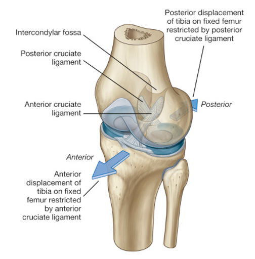
| anterior cruciate ligament |
internal to joint capsule; frequently injured in sports injuries | |
| posterior cruciate ligament |
internal to joint capsule |

Note that what we are calling the patellar tendon is really a combination of both tendon and ligament. Its upper part extends from the quadriceps muscles, while its lower part extends between the bone of the patella and the bone of the tibia. The knee is stabilized by two external ligaments (the tibial collateral ligament and the fibular collateral ligament) and two ligaments that are inside the joint capsule (the anterior cruciate ligament and the posterior cruciate ligament). The cruciate ligaments are so named because they cross each other.
The articular cartilage is represented on the model as the smooth and shiny layer at the end of the bone and is easiest to see on the end of the femur. In addition to articular cartilage, the knee joint contains two fibrocartilage pads called the menisci (singular: meniscus). Knowing that the fibula is the lateral bone enables you to distinguish the lateral meniscus from the medial meniscus.
The figure below illustrates the structure of the menisci.
The cruciate ligaments help to prevent the femur from sliding forwards or backwards on the tibia.
Optional videos
These videos from Acland’s Video Atlas of Anatomy will help you understand the relationship between the cartilage, ligaments, and fibrous capsule of the knee joint.
2.2.2 Cartilages and cruciate ligaments of the knee joint
2.2.3 Collateral ligaments of the knee joint, patellar tendon, quadriceps bursa, joint capsule