
11 Bones of the Upper Limb
Anatomical Terms
Before beginning to study anatomy of the bones, it is important to define some anatomical terms. When studying anatomy, it is important to have terms of direction that specify the relative positions of structures. Below is a list of the terms of direction we will use in this class. They are given in pairs because they define opposite directions along a particular axis.
Anatomy Terms of Direction
- anterior/posterior
- ventral/dorsal
- medial/lateral
- superior/inferior
- cranial/caudal
- proximal/distal
The terms of direction are illustrated in the figure below.
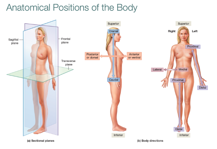
Another useful definition is anatomical position. The person in the figure above is drawn in anatomical position. In anatomical position, the limbs are extended straight and the palms face anteriorly. Anatomical position is a reference position that can be used to define limb movements. We will use this along with the sectional planes to define the actions of the muscles. Anatomical position is also key in defining the medial and lateral positions of the bones in the forearm (lower arm).
In addition to the terms of direction, it is useful to know the meaning of certain terms used to describe features on the bones. Muscle attachment points create rough spots where the tendon knits into the bone. In the joint, cartilage on the surface of the bone (articular cartilage) creates a smooth region known as a condyle. Below are definitions of terms we will use.
Glossary of Terms in Bone Anatomy
process: a projection that sticks out from the bone
tuberosity: a rough raised portion of bone where tendon of muscle attaches
condyle: smooth region that forms a joint with another bone; in life, covered with articular cartilage
epicondyle: a small projection above a condyle
head: proximal rounded extension of bone, may be separated from the shaft of the bone by a neck
articulation: joint
diaphysis: shaft of a long bone
epiphysis: end of the bone; growth plate is located in the epiphysis of growing bones
Using features of the bones and the terms of direction, you will learn how to distinguish whether a bone is from the left or right side of the body.
Bones of the Upper Limb
For reference, the figure below shows how all the bones of the upper limb fit together.
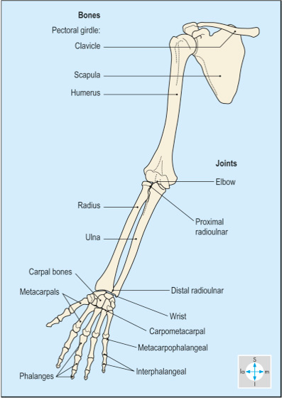
The arm is attached to the trunk by the pectoral girdle, or shoulder girdle. The bones of the shoulder girdle are the clavicle and scapula.
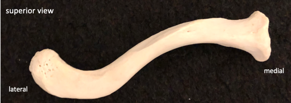
The figure below shows a superior view of the clavicle.
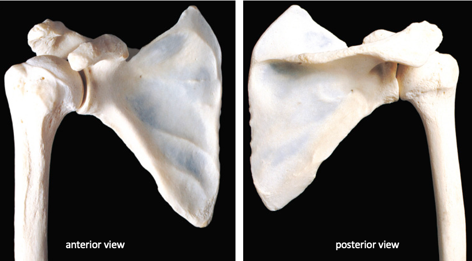
The figure below shows anterior and posterior views of the scapula. The large ridge on the posterior side of the scapula is called the spine. The glenoid cavity is the shallow depression on the lateral side of the bone that articulates (forms a joint with) the head of the humerus.
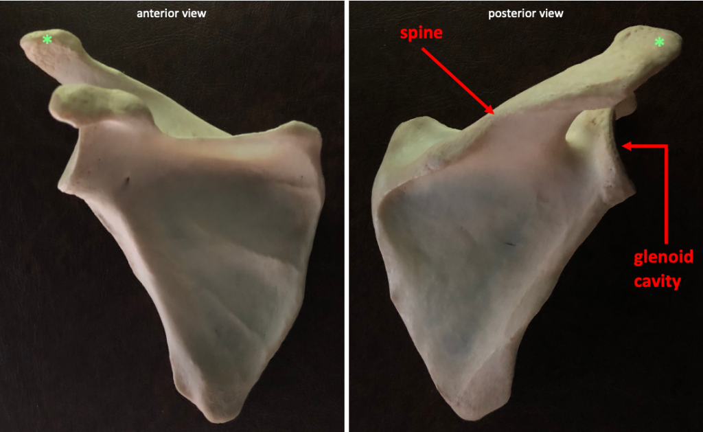
This lateral view shows the surface of the glenoid cavity.
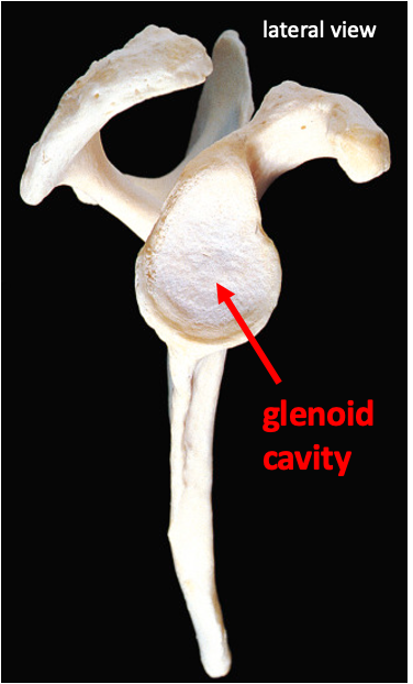
In life, the glenoid cavity is covered with articular cartilage. Because the glenoid cavity is so shallow, there is also a small ridge of fibrocartilage around the edge (called the glenoid labrum), which helps to stabilize the humerus.
The figure below shows how the humerus fits together with the bones of the shoulder girdle.
The humerus is the bone of the upper arm. The rounded head of the humerus is a key feature that tells you which is the medial side and proximal end of the bone.
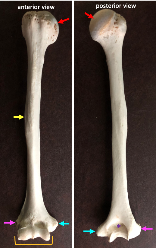
The distal end of the humerus articulates with the two bones of the forearm (lower arm): the radius and the ulna. The condyle on the distal humerus has a complex shape. Above the condyle are the medial epicondyle and the lateral epicondyle. The medial epicondyle is a fairly large projection. This is what we call the “funny bone”: the ulnar nerve wraps around the medial epicondyle. If you hit this part of the bone, you will feel pain in the medial part of your lower arm.
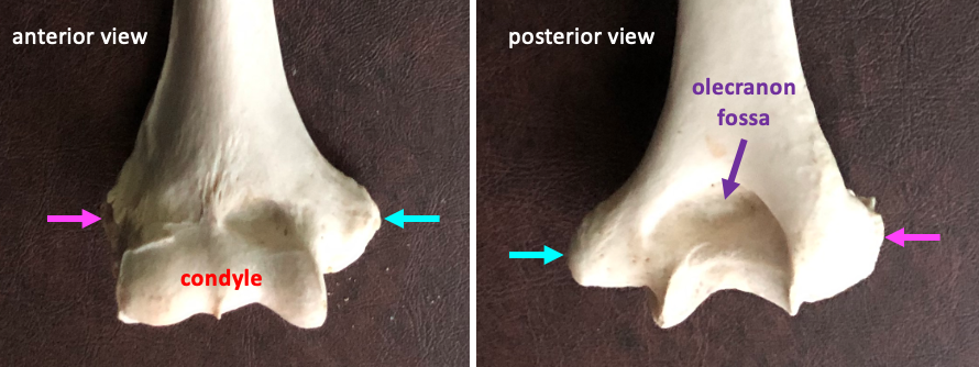
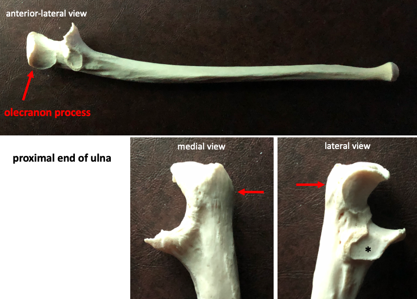
The olecranon fossa is a depression located on the posterior side of the distal humerus. This depression is where the olecranon process of the ulna fits.
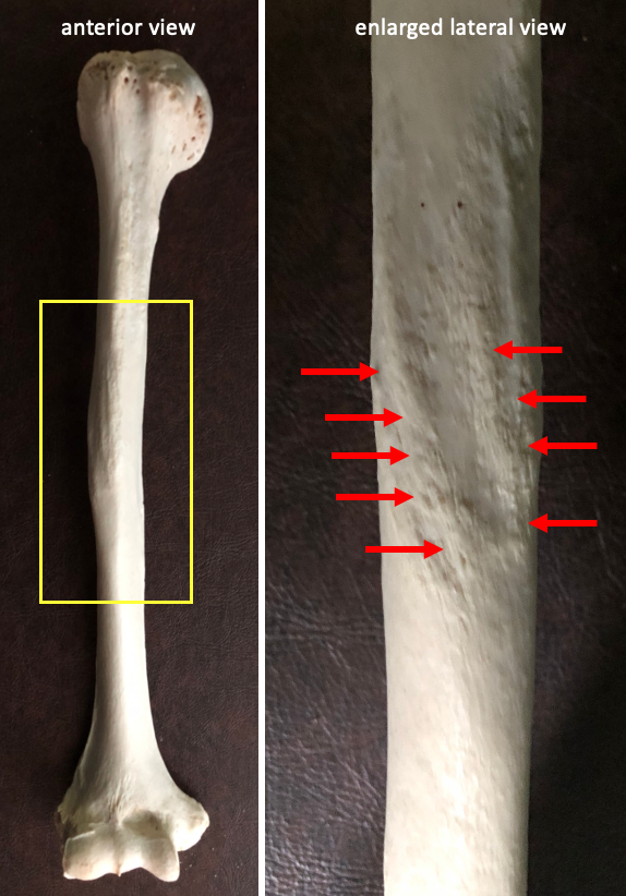
The deltoid tuberosity is a V-shaped rough area on the lateral surface of the humerus. This is the insertion (distal attachment) for the deltoid muscle.
The two bones of the forearm are the radius and the ulna. When the arm is in anatomical position (palm facing forward), the radius is lateral and the ulna is medial.
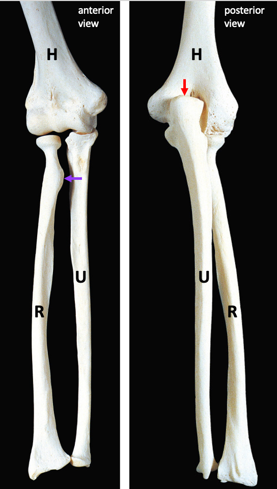
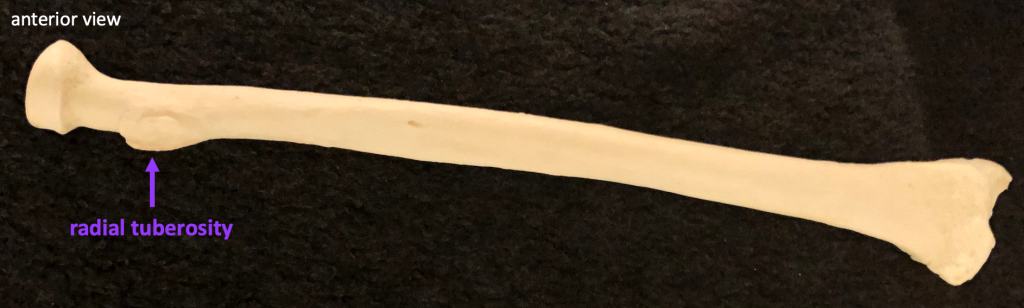
The radial tuberosity is a rough projection near the proximal end of the radius. This is the insertion for the biceps brachii muscle.
The olecranon process is the large curved proximal end of the ulna. This forms the bony part of the elbow.
Optional videos
Below are links to videos in the Acland’s Video Atlas of Anatomy. They are helpful in allowing you to get a sense of the bones in three dimensions. They also will introduce you to the movements around the joints, which we will be studying in our class about muscle anatomy. Video links below open in a new tab.
1.1.2 The clavicle and scapula
1.1.4 The shoulder joint and its movements
1.2.2 Bones of the arm and forearm