
10 Optional: Autonomic Control of Pupil Diameter in the Eye
The pupil is the hole in the center of the iris that allows light to get into the eye. The diameter of the pupil is controlled by smooth muscles in the iris of the eye. The pupil is in the center of the eye and appears black, while the iris is the colored portion that surrounds it. Autonomic control of pupil diameter is a classic example where the sympathetic and parasympathetic divisions have antagonistic actions.
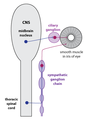
Refer to the figure above. Sympathetic preganglionic neurons located in the thoracic spinal cord activate sympathetic postganglionic neurons in the sympathetic ganglion chain. The sympathetic postganglionic neurons release NE, which stimulates contraction of the radial muscles in the iris and causes the pupil to dilate(get larger). The parasympathetic preganglionic neurons are located in a nucleus in the midbrain. They travel via a cranial nerve (the occulomotor nerve) to activate postganglionic neurons in the ciliary ganglion, which is located just behind the eye. Parasympathetic postganglionic neurons release ACh, which stimulates contraction in circular muscles in the iris and causes the pupil to constrict (get smaller).