
9 Peripheral Nervous System
Introduction
The central nervous system (CNS) consists of the brain and spinal cord; everything outside of the dura mater is considered the peripheral nervous system (PNS). The PNS can be divided into two major divisions based on the way that information flows. The afferent division consists of neurons that are bringing sensory information about the periphery toward the CNS, while the efferent division consists of neurons that are conveying information away from the CNS, and out to control muscles and organs in the body. The efferent division is further divided into the somatic efferent division, which consists of neurons that control skeletal muscles, and the autonomic efferent division, which consists of the neurons that control all other organs (collectively termed viscera).
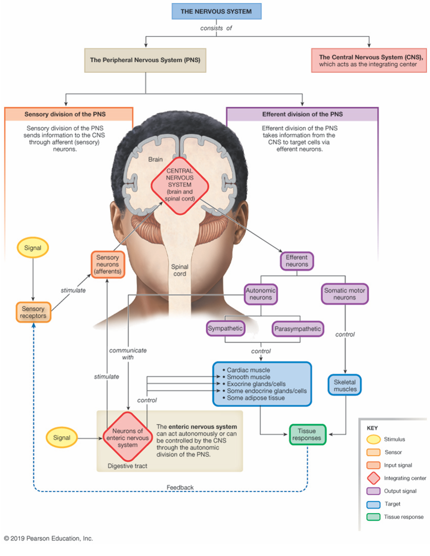
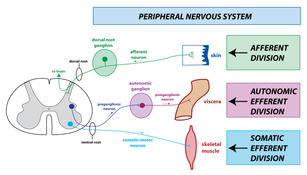
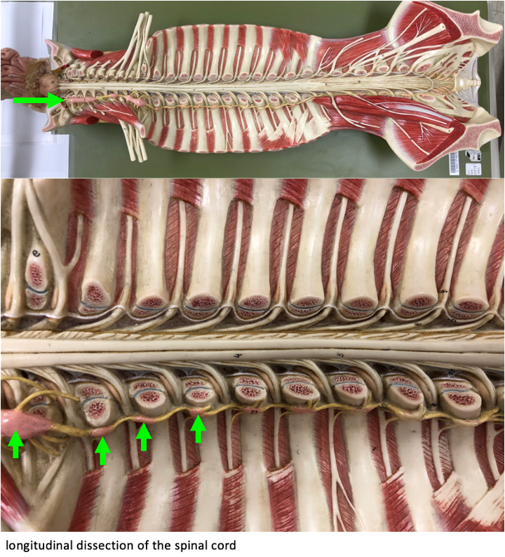
In the previous chapter, we looked at the anatomy of the peripheral nerves and their association with the spinal cord. Spinal nerves are bundles of axons containing both afferent and efferent axons. But close to the spinal cord, the axons are separated into dorsal roots (afferent axons) and ventral roots (efferent axons). In this chapter, we will look at histology, and focus on identifying specific types of neurons in the peripheral nervous system. Below is a figure that provides an overview of the three divisions of the peripheral nervous system.
Afferent Division
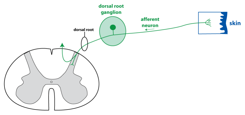
Afferent neurons have a unique structure that differs from most other neurons. Their dendrites (known specifically as sensory dendrites) are located out in the periphery (many are in the skin). The sensory dendrites are directly attached to a long axon that projects into the dorsal spinal cord via the dorsal root. The cell bodies of afferent neurons are clustered around the dorsal root in the dorsal root ganglion. The large, round cell bodies of afferent neurons are attached to their axons by a short stalk.
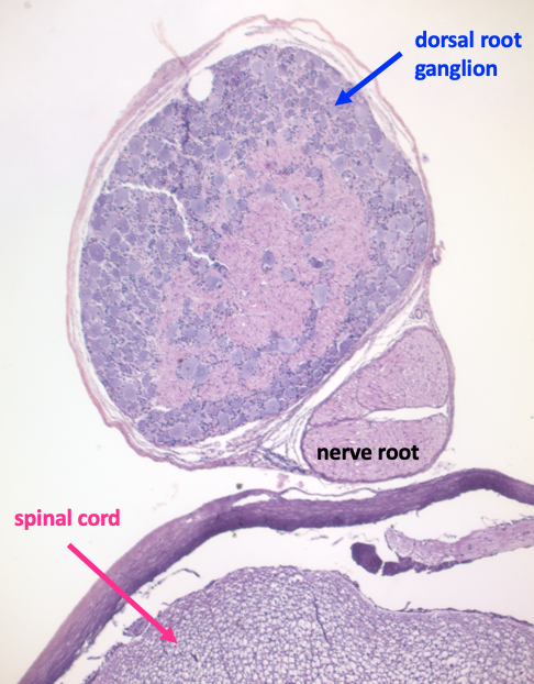
Below is a low magnification view of a dorsal root ganglion (top) adjacent to the spinal cord (bottom). Just visible at this magnification are the large cell bodies of afferent neurons (purple circles). The pink material centrally is axons of the dorsal root.
The next image shows a high magnification view of the dorsal root ganglion. The pink circles are axons of the dorsal root. The large, pale purple circles are the cell bodies of the afferent neurons. The small dark nuclei that surround the neuronal cell bodies are the nuclei of satellite cells, a type of glial cell found in the dorsal root ganglia.
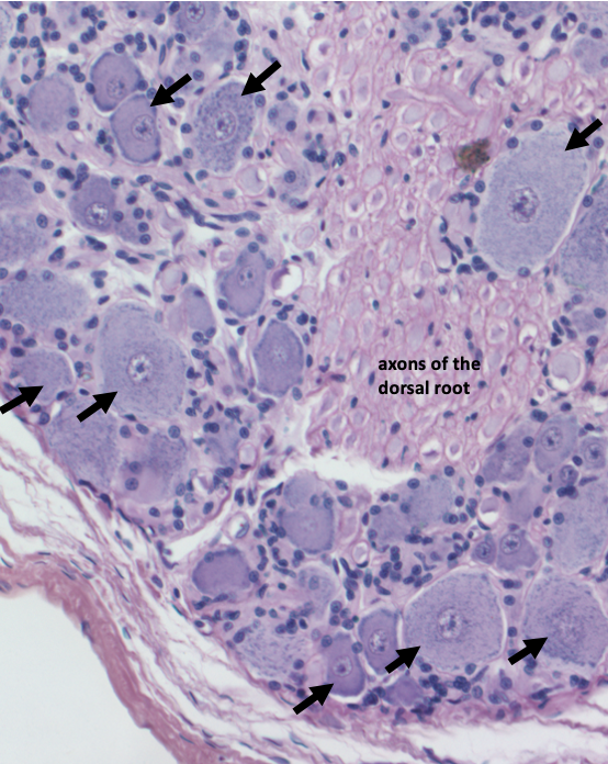
Afferent neurons are large cells compared to other cells (compare the neuronal nuclei with the satellite cell nuclei, for instance). Besides having a large cell body, the nucleus of a neuron contains a prominent nucleolus. This is the organelle located within the nucleus that is responsible for producing ribosomal RNA. A prominent nucleolus is a characteristic of cells that engage in a lot of protein synthesis. (Note that a nucleus and a nucleolus are not visible in every neuronal cell body; because this is a thin slice of tissue, and sometimes the plane of section misses certain elements in a large cell.)
The sensory dendrites in the periphery are the part of the afferent neuron where sensory transduction occurs. The sensory dendrites contain gated ion channels that determine the modality of the afferent neuron (touch, pain, temperature). For example, touch-sensitive neurons contain mechanically-gated ion channels. In response to touch, these channels open, converting the mechanical stimulus into a change in membrane potential that is called the receptor potential.
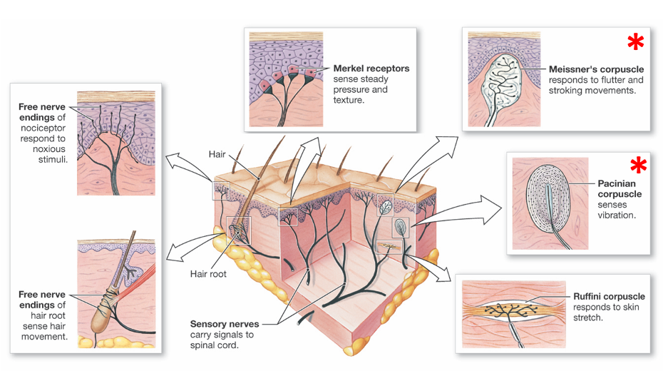
Sensory dendrites may exist as free nerve endings, or they may be encapsulated in specialized connective tissue structures. Free nerve endings are difficult to see in histology slides without special staining mechanisms. However two types of encapsulated touch sensors are readily visible in histological sections of skin: Pacinian corpuscles and Meissner’s corpuscles.
Pacinian Corpuscle
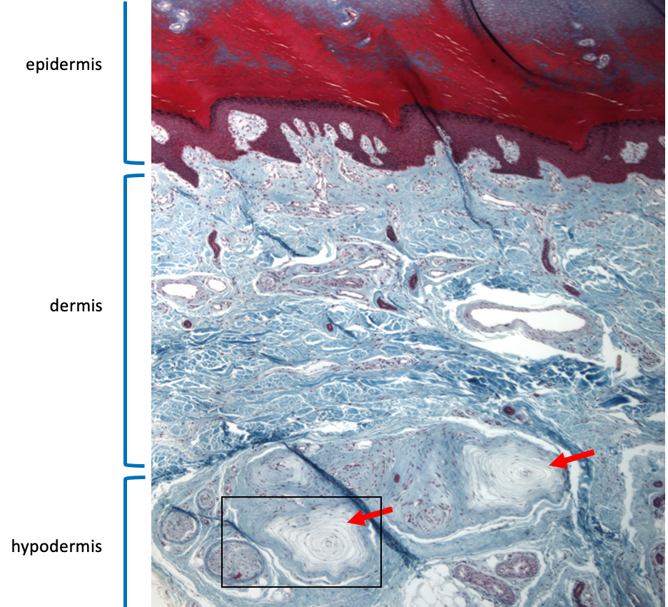
Pacinian corpuscles are located in the hypodermis (also known as the subcutaneous layer) of the skin. Their sensory dendrites are encapsulated in concentric layers of connective tissue, so they look a bit like onions. The afferent neurons associated with Pacinian corpuscles are rapidly adapting and they respond best to vibration.
In the low magnification view below, two Pacinian corpuscles can be seen in the hypodermis. Just to the left of the Pacinian corpuscles, there are several small nerves cut in cross section. The box highlights the region that is enlarged in the next figure.
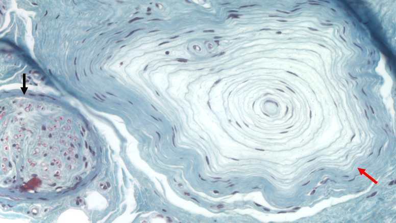
The high magnification view below allows you to see the concentric layers of connective tissue in the Pacinian corpuscle.
Meissner’s Corpuscle
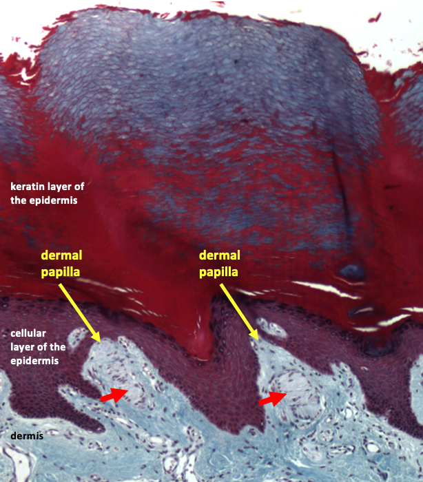
Meissner’s corpuscles are located in the upper part of the dermis, just under the epidermis. Meissner’s corpuscles sense fine touch. The section of skin in the figures below is taken from the palmar surface of the fingertip (the side where the fingerprint is). Meissner’s corpuscles are more abundant in this skin, making it much more sensitive to fine touch.
In the low magnification view, two Meissner’s corpuscles are visible in the part of the dermis that protrudes into the epidermal layer (called a dermal papilla).
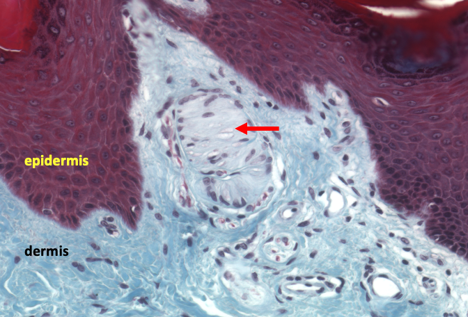
The next picture is a high magnification view of the dermal papilla on the right. The Meissner’s corpuscle is the grayish structure in the center of the papilla. The sensory dendrites are embedded in the layers of connective tissue in the Meissner’s corpuscle.
Somatic Efferent Division
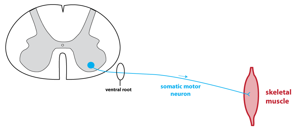
The somatic efferent division consists of the neurons that innervate skeletal muscle. These somatic motor neurons have their cell bodies in the ventral horn of the spinal cord. Their axons leave the spinal cord via the ventral root. They synapse onto skeletal muscle cells, where they release the neurotransmitter acetylcholine. The acetylcholine acts on nicotinic acetylcholine receptors to cause an excitatory post synaptic potential. The synapse between a somatic motor neuron and a skeletal muscle fiber is called the neuromuscular junction.
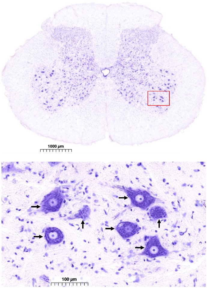
The figure below shows a section of the spinal cord at the sacral level. A section from the sacral level will have lots of somatic motor neuron cell bodies since this region of the spinal cord innervates the legs.
This tissue is treated with the Nissl stain, which highlights neuronal cell bodies. The top image is a low magnification view which shows the gray matter, and indicates the region in the ventral horn where somatic motor neuron cell bodies are located. The bottom part of the figure focuses in on several somatic motor neurons.
Autonomic Efferent Division
The autonomic division provides innervation to all other tissues that receive neural control. Targets of the autonomic nervous system include cardiac muscle in the heart, smooth muscle in organs and blood vessels, and secretory cells in the digestive tract.
The organization of the autonomic efferent division is more complex than the somatic efferent division. Information must flow through two neurons as it travels to a target: a preganglionic neuron and a postganglionic neuron. The figure below shows this relationship.
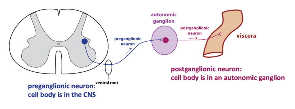
The preganglionic neuron has its cell body in the central nervous system, with its axon in the ventral root. The axon travels to an autonomic ganglion, where it forms a synapse with a postganglionic cell. Postganglionic neurons synapse with target cells.
Another complexity is that there are two main autonomic divisions: the sympathetic division and the parasympathetic division. Often a target will receive inputs from both sympathetic and parasympathetic neurons (dual innervation), with each division causing an opposite effect. A useful organizing principle is to remember that sympathetic activity is activated during acutely stressful situations (the “fight-or-flight” response). For example, activation of the sympathetic nervous system causes an increased heart rate, dilation of the pupils, and diversion of blood flow to skeletal muscles. By contrast, when the body is “resting and digesting”, parasympathetic nervous system activity dominates. Parasympathetic input reduces heart rate and promotes digestive processes.
There are also key differences in the anatomical organization of the sympathetic and parasympathetic divisions. The cell bodies of parasympathetic preganglionic neurons are located in the brainstem and the sacral spinal cord. Parasympathetic preganglionic neurons have long axons because parasympathetic ganglia are located close to targets.
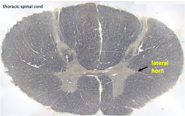
The cell bodies of sympathetic preganglionic neurons are located in the lateral horn of the thoracic spinal cord.
The sympathetic chain ganglia are a string of ganglia located close to the spinal cord. The cell bodies of sympathetic postganglionic neurons are found in the sympathetic chain ganglia.
Finally, the sympathetic and parasympathetic postganglionic neurons release different neurotransmitters at target tissues. We will focus on this aspect of autonomic nervous system physiology in lecture, but you will probably find the description below useful. Sympathetic postganglionic neurons release the neurotransmitter norepinephrine (abbreviated NE)*. NE acts on adrenergic receptors. Parasympathetic postganglionic neurons release the neurotransmitter acetylcholine (abbreviated ACh)*, which acts on muscarinic receptors. Both adrenergic and muscarinic receptors are G-protein coupled receptors (GPCRs) The significance of this information is that drugs targeting these receptors are used to affect autonomic activity.
| postganglionic neurotransmitter | receptor type on target cell | |
| SYMPATHETIC | norepinephrine | adrenergic |
| PARASYMPATHETIC | acetylcholine | muscarinic |
*There are a few exceptions to the rule that sympathetic postganglionic neurons release NE and that parasympathetic postganglionic neurons release ACh. In physiology, it seems there are always exceptions to every rule.