8 Spinal Cord Anatomy
Spinal Cord Tissue
The most important terms of direction for studying spinal cord anatomy are ventral (which means “towards the stomach”) and dorsal (which means “towards the back”). In human anatomy, with our upright posture, the ventral side corresponds to the anterior side, and the dorsal side corresponds to the posterior side.
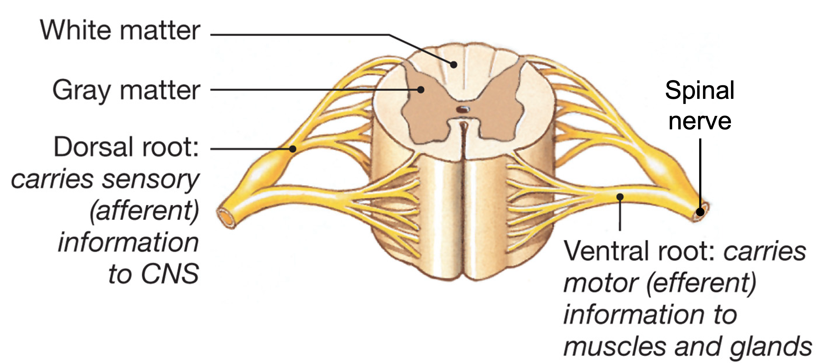
Like the brain, spinal cord tissue is divided into gray matter, containing primarily neuronal cell bodies, and white matter containing axons, arranged into tracts. The gray matter of the spinal cord is located centrally and has roughly the shape of a butterfly. The white matter is around the outside. The central canal, at the center of the gray matter, is a space containing cerebrospinal fluid that is continuous with the ventricles in the brain.
A further degree of organization is that there is a separation of afferent information, and efferent information. Afferent neurons (afferent means “conducting towards”) convey sensory information from the periphery into the central nervous system. The axons of afferent neurons enter the spinal cord on the dorsal side. They form synapses with cells in the dorsal horn, which is the term for the narrowish region of grey matter that extends out to the edge of the spinal cord. Efferent neurons (efferent means “conducting away from”) have their cell bodies in the spinal cord, and extend axons out to the periphery to control muscles and other cells. In particular, somatic motor neurons that innervate skeletal muscle cells have their cell bodies located in the ventral horn. Further away from the spinal cord, afferent and efferent axons are bundled together in spinal nerves that travel to particular locations (for instance, your fingertip).
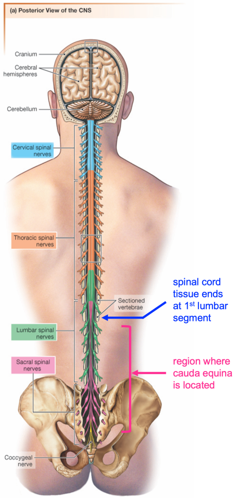
Segments of the spinal cord are divided into 4 basic regions (cervical, thoracic, lumbar, and sacral), named for the level at which the spinal nerves exit the vertebral column. The size and shape of the gray matter varies depending on the segment of spinal cord. This is because the segments that innervate the arms and legs have many more neurons, and so have correspondingly larger dorsal and ventral horns.
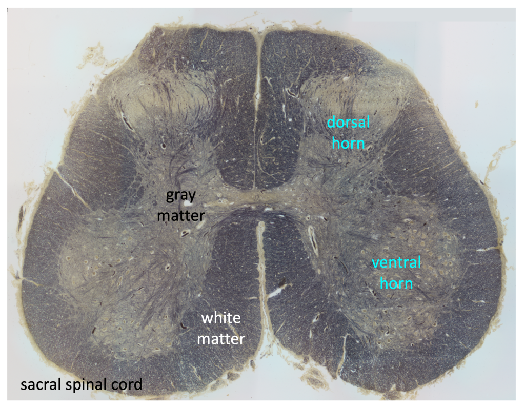
This first micrograph shows a section from the sacral region of the spinal cord. The tissue is stained with cresyl violet, a stain that binds to rough endoplasmic reticulum and so stains the neuronal cell bodies. The butterfly-shaped gray matter is darker than the outer white matter. The central canal is visible at the center of the gray matter. The central canal is a space filled with cerebrospinal fluid and is continuous with the ventricles of the brain.
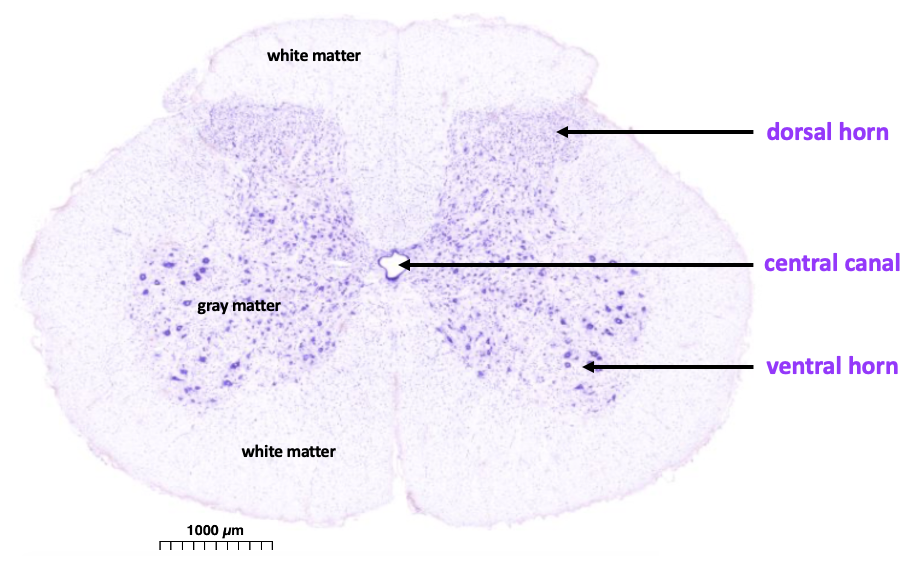
https://histologyguide.com/slideview/UCSF-163-spinal-cord/06-slide-1.html
The next figure shows a section of spinal cord from the thoracic level. The tissue is stained with a dark substance that binds to membranes and so the white matter (which contains many myelinated axons) stains dark gray.
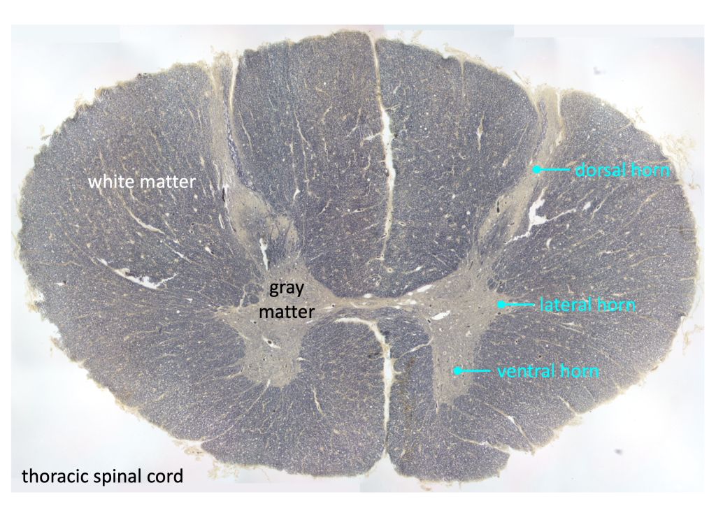
Neurons at the thoracic level innervate skin, organs, and muscles in the trunk. The dorsal horn is narrow and extends out to the edge of the spinal cord, while the ventral horn is small and rounded. The thoracic region is notable for the presence of the lateral horn, which is the location of the cell bodies of sympathetic preganglionic neurons.
Below is a similarly stained section from the sacral region. Notice the much larger size of the ventral horn, which is full of somatic motor neuron cell bodies innervating skeletal muscles in the legs. The dorsal horn is larger too, because there is relatively more sensory information coming from the legs than from the trunk.
Nerves, Roots, Dorsal Root Ganglia
We will use spinal cord models to learn about the anatomy of the nerves and roots. The model shown in the picture below is a cross section of the cervical spinal cord.
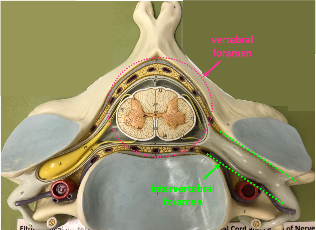
The spinal canal is created by the successive vertebral foramina, the vertebral foramen being the large space within each vertebra. At the side, is a space called the intervertebral foramen, which contains the dorsal and ventral roots and the dorsal root ganglion.
We looked at this model in the chapter about ventricles and CSF. Follow this link to review those structures.
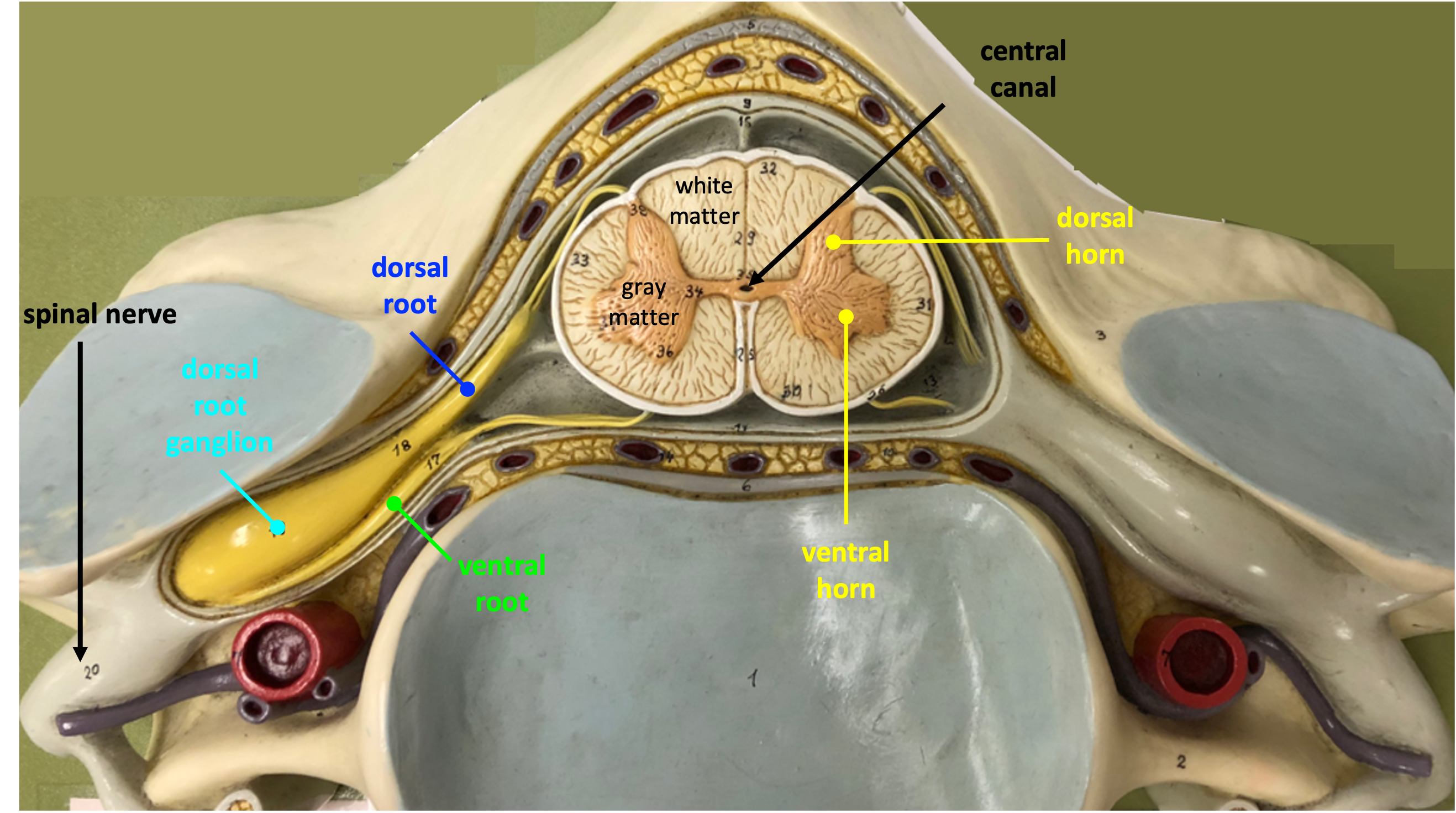
The spinal nerve contains both afferent (sensory) axons and efferent (motor) axons in one bundle. Close to the spinal cord, the afferent and efferent axons are found in separate bundles called the dorsal root and ventral root.
The dorsal root ganglion is the swelling that is found along the dorsal root. The dorsal root ganglion contains the cell bodies of afferent neurons (see the next chapter on the organization of the peripheral nervous system). The dorsal root ganglia are located just outside the dura mater. Note that the dorsal root ganglion does not lie in a position that is dorsal to the spinal cord, but rather gets its name because it is a cluster of neuronal cell bodies (“ganglion”) that surrounds the dorsal root. The cell bodies of neurons whose axons are found in the ventral root are located in the spinal cord. You should also be able to distinguish gray matter and white matter in this model and be able to identify the central canal, dorsal horn, and ventral horn.
The other model shows a longitudinal dissection of the spinal cord. The model is set up as if the cadaver was lying on its back, with all of of the tissues ventral to the spinal cord removed. The dura mater has been cut. Thus, we are looking at the ventral surface of the spinal cord.
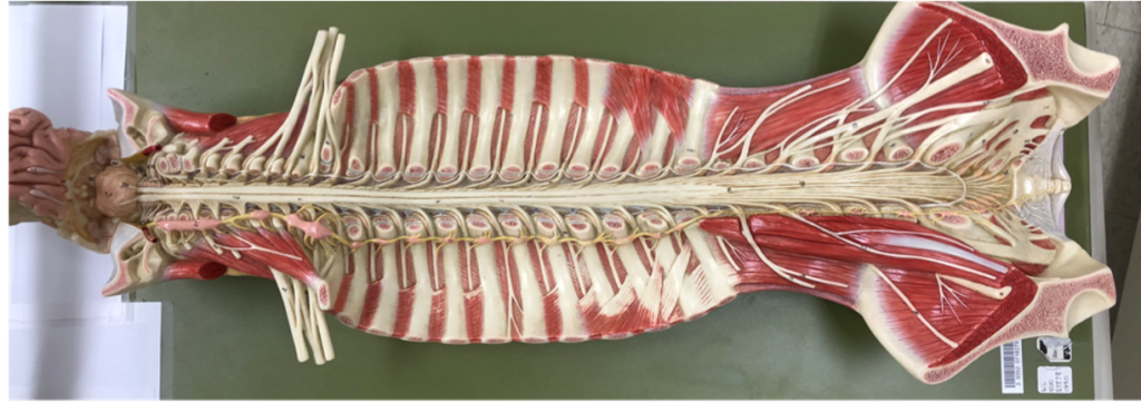
In the full view of the model above, notice how large the spinal nerves are that supply the arms and the legs (the spinal nerves are white structures leaving spinal canal in each segment). In these regions, the spinal nerves divide and rejoin to form a network called a plexus. The more superior plexus whose nerves innervate the arms is called the brachial plexus; the lower one whose nerves innervate the legs is called the lumbosacral plexus.
In a close-up view, one can see that on one side of the model (the top side of the picture below), the roots are cut, revealing the dorsal roots. The ventral roots are visible on the surface on the undissected side (bottom side of the picture below).
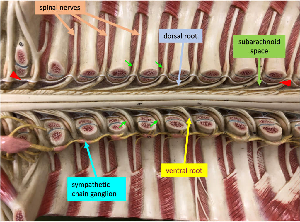
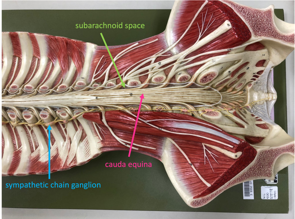
On the model’s right side, you can see the sympathetic chain ganglia (there are sympathetic chain ganglia on both sides of the body, but they are only modeled on the right side in this particular model). These ganglia contain the cell bodies of sympathetic postganglionic neurons. In the model the sympathetic chain ganglia are bright pink and connected by yellow processes.
The dura mater has been cut to reveal the spinal cord. The white wavy line represents the cut edge of the dura mater on this model; thus the space adjacent to the spinal cord is the subarachnoid space.
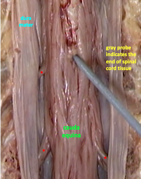
The dorsal and ventral roots travel some distance caudally before they join to form a spinal nerve that exits the vertebral canal. These distances become longer for more caudal segments. The tissue of the spinal cord only extends to the beginning of the lumbar region, so the most caudal part of the vertebral canal is a group of long roots known as the cauda equina (because it resembles a horse’s tail).
The cauda equina is visible in a close-up of the caudal region of the model.
A spinal tap is when a needle is inserted into the subarachnoid space in order to take a sample of the cerebrospinal fluid. This is done in the region of the cauda equina in order to reduce the risk of damage to the spinal cord.
Videos from Acland’s Video Atlas of Human Anatomy
The Acland’s Video Atlas of Anatomy provides narrated video demonstrations using high quality cadaver dissections. These videos contain more detail and terminology than I expect you to learn for this class. In each case, focus on being able to identify the terms given in purple boldface. Dr. Acland speaks with a British accent, so you may want to utilize the closed captions. A pdf transcript is also available for each video.
There are two videos about spinal cord anatomy that are useful for this class. The first video (video 3.1.8) shows a cross section of the spinal column. This video shows very nicely the relationship of the three layers of meninges. A link to this video can also be found in the chapter: Ventricles, Cerebrospinal Fluid, and the Blood-Brain Barrier. In video 3.1.8, be able to identify:
spinal cord
dura mater
subarachnoid space
epidural space
3.1.8 Spinal cord (cross-section), spinal meninges, dural sac
The second video (video 3.1.9) is a posterior (dorsal) view of the spinal cord. This video shows the end of the spinal cord tissue and the cauda equina. It also shows how the “filaments” (nerve rootlets) leaving the dorsal and ventral sides of the spinal cord coalesce to form the dorsal and ventral roots, and then how the roots come together to form a spinal nerve. In video 3.1.9, be able to identify:
spinal cord
cauda equina
dorsal root
ventral root
spinal nerve
dorsal root ganglion
3.1.9 Spinal cord (from behind), nerve roots
The figure below is a key screenshot from video 3.1.9 showing the caudal equina.