5 Nervous System Histology
Structure of Neurons
The information-carrying cells in the nervous system are called neurons. Neurons are unique cells in that they extend long processes (sometimes more than a meter in length) and make direct connections, called synapses, with their target cells. Below is a schematic drawing illustrating the different parts of a neuron. Be able to identify the different parts of a neuron that are highlighted in purple boldface.
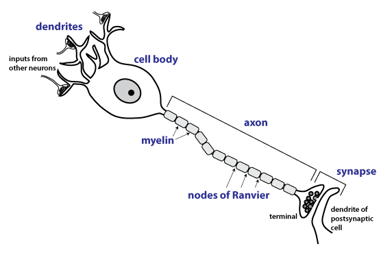
The dendrites of a neuron constitute an input region where a neuron receives synapses from other neurons. For neurons that sense touch, temperature, and pain in the skin (afferent neurons) the input region consists of sensory dendrites that contain specialized ion channels that open in response to the particular sensory modality. For instance the sensory dendrites of touch-sensitive neurons have mechanically-gated ion channels.
Signals from multiple inputs are integrated in the neuronal cell body (also called the soma of the neuron). The cell body is also the metabolic center of the neuron. Neurons can be very large cells with axons that extend long distances, and so need a large cell body. Neurons are often actively synthesizing lots of protein so they have an extensive network of rough endoplasmic reticulum (rough ER) and a large and prominent nucleolus, which is the organelle inside the nucleus where ribosomal RNA is made.
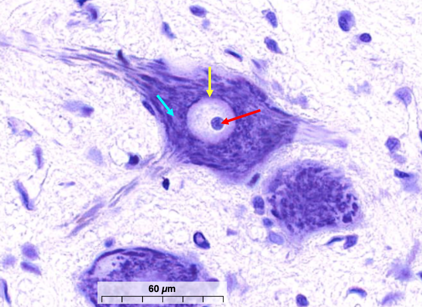
The figure below shows a section from the sacral spinal cord, stained with cresyl violet, which is a basic dye that highlights neuronal cell bodies. In the spinal cord, the gray matter (the region containing neuronal cell bodies) is found centrally. The outer pale-staining tissue contains bundles of axons and is called the white matter. The ventral region of the spinal cord contains many large cell bodies of somatic motor neurons, the neurons that directly excite skeletal muscle cells.
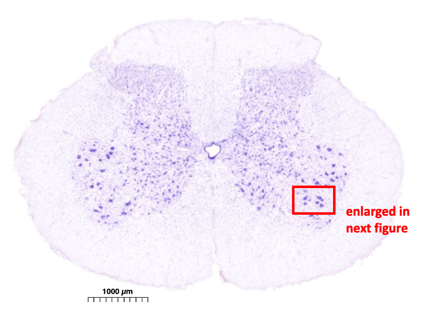
https://histologyguide.com/slideview/UCSF-163-spinal-cord/06-slide-1.html
This next figure focuses in on the ventral region of the gray matter, which is called the ventral horn. The cell bodies of seven somatic motor neurons are visible.
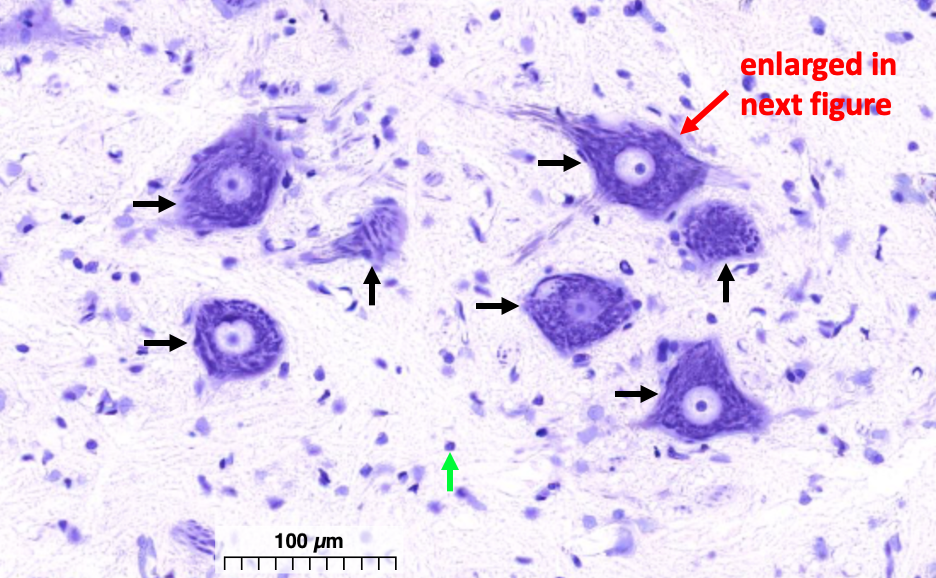
The cell body shows characteristic purple/blue staining of the rough ER. Inside the nucleus is the darkly stained nucleolus. These features are highlighted in the next figure.
The output region of the neuron consists of the axon and axon terminal, which forms a synapse with a target cell. The axon conducts action potentials and thus contains voltage-gated ion channels in its plasma membrane. Action potentials are rapid, all-or-nothing electrical signals that can conduct long distances without decrement. This is necessary because some axons can be up to a meter long.
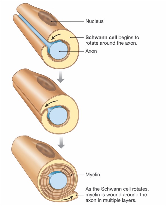
Myelin
Many axons are myelinated. Myelination speeds axonal conduction. In the central nervous system, myelin is formed by oligodendrocytes. In the peripheral nervous system, myelin is formed by Schwann cells. Myelin consists of many tightly wrapped concentric layers of plasma membrane.
The highly precise structure of myelin is visible in the following electron micrographs. The first one shows a cross-section of a myelinated axon in the peripheral nervous system.
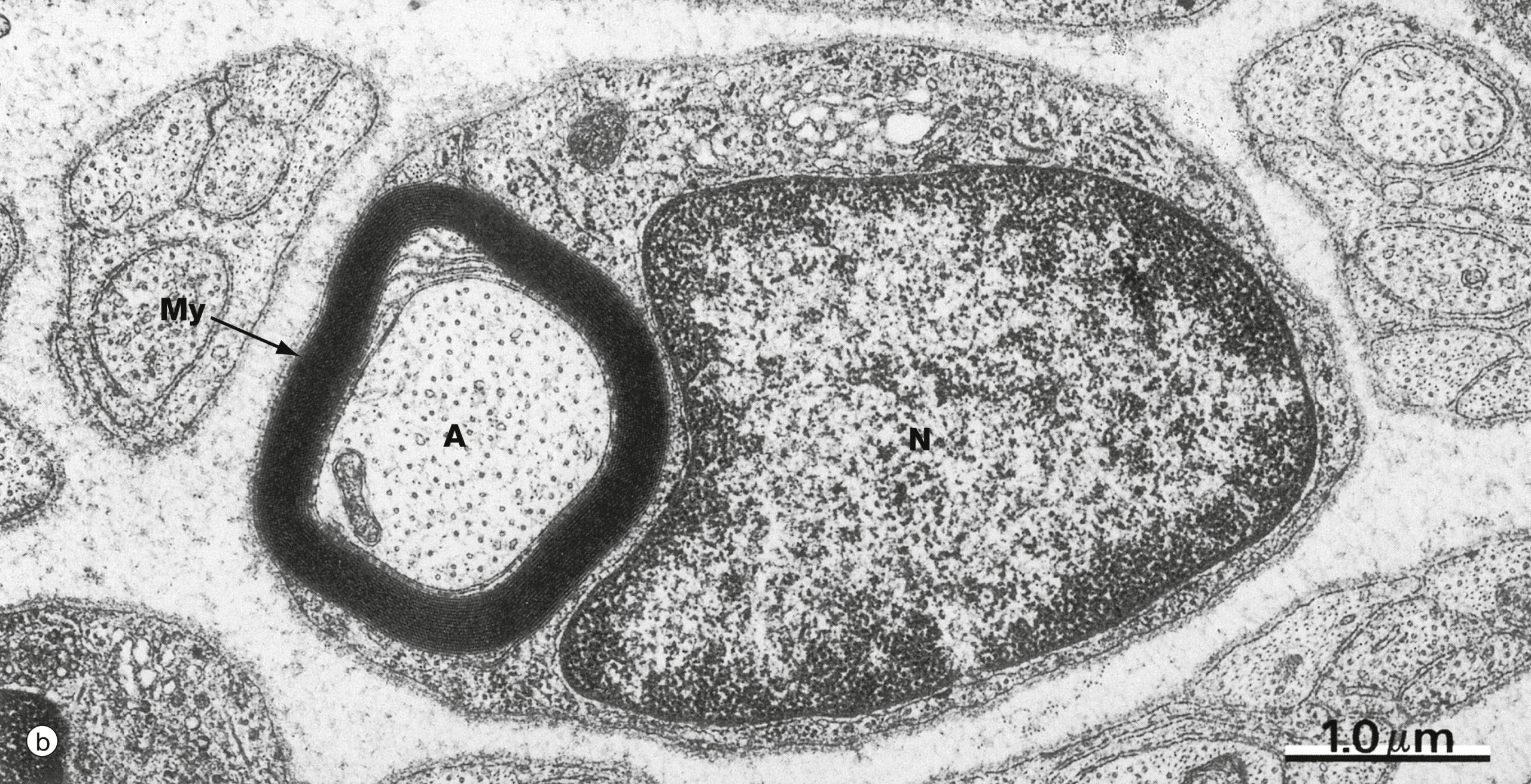
The small dots visible in the axon are microtubules, which are used in transporting organelles and proteins along the length of the axon.
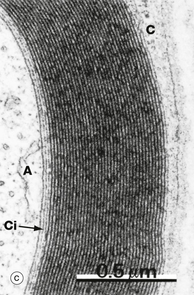
The next micrograph is a highly-magnified view of myelin. The Schwann cell plasma membranes fuse together and all the cytoplasm is extruded to form the highly compacted myelin sheath.
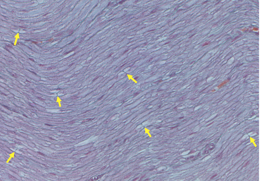
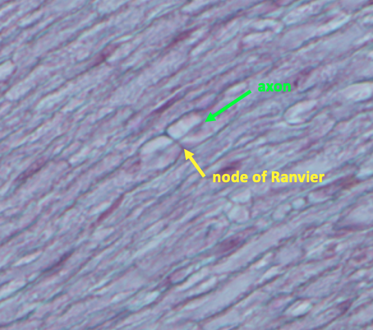
Myelin forms in bundles covering short segments of the axon; there is a bare, unmyelinated stretch of membrane between two bundles of myelin that is called a node of Ranvier. The voltage-gated ion channels that generate the action potential are concentrated at the nodes of Ranvier.
The next two figures show a longitudinal section of a peripheral nerve. The axons are running across the view from right to left. During histological processing, the lipid is mostly dissolved, so the bundles of myelin look like empty bubbles. When the section passes through a node of Ranvier, it appears as a line running perpendicular to the axon. Several nodes are indicated by yellow arrows.
Synapses
The synapse is where a neuron communicates with its target cells. In the central nervous system, a single cell typically makes tens of thousands of synapses.
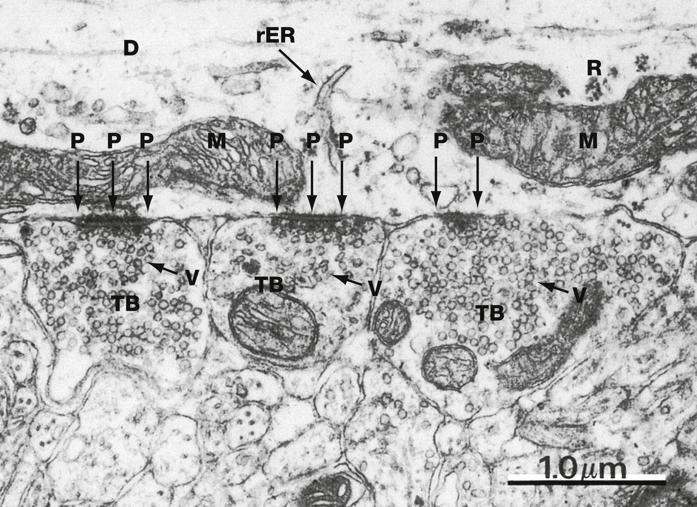
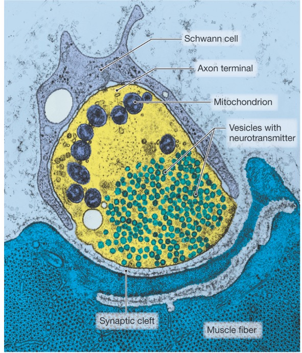
The synapse consists of the axon terminal of the presynaptic cell, and a specialized region of the postsynaptic cell. There can be electrical synapses, where intercellular channels called gap junctions allow electrical current to flow between cells. The predominant type of synapse in the nervous system is a chemical synapse in which the presynaptic cell releases a neurotransmitter into a narrow space called the synaptic cleft. The neurotransmitter diffuses across the synaptic cleft and to its receptor on the postsynaptic cell. Neurotransmitters are packaged in synaptic vesicles and released through exocytosis. In an electron micrograph, a chemical synapses can be recognized by the presence of synaptic vesicles.
The first figure shows a pseudo-colored electron micrograph of the neuromuscular junction, which is the synapse between a somatic motor neuron and a skeletal muscle cell. This synapse is unique in that it is very large. This large synapse guarantees that each time the somatic motor neuron fires an action potential it will activate a contraction in the skeletal muscle cell.
In contrast to the neuromuscular junction, the much smaller synapses in the CNS mean that a postsynaptic cell will need to add together the inputs from many cells before it is excited to fire an action potential. The figure below shows three axon terminals forming chemical synapses in the CNS. Synaptic vesicles can be seen in the axon terminals, which are marked TB (for terminal bouton).