
1 Lab 1 External brain morphology
Specific Learning Objectives
- Describe and locate the three meningeal layers surrounding the brain; identify and describe the dural reflections and CNS cisterns
- Relate features of the brain’s surface to the cranial fossae of the skull
- Describe and locate the major parts of the forebrain, midbrain and hindbrain visible in an intact brain and in a mid-sagittally sectioned brain
- Locate and identify the lobes of the brain, and major sulci and gyri, in superior, inferior, lateral, and medial (sagittal) views of the whole brain, and describe the functions of the lobes
- Correlate the major functional areas of the cerebral cortex with anatomic landmarks
- Describe the basic function of and locate the cerebellum
- Distinguish the three parts of the brainstem (midbrain, pons, medulla) based on their location and surface morphology
- Locate the cranial nerve attachment points and summarize the function of each of the cranial nerves
- Locate and identify the external features of the spinal cord, including posterior and anterior roots, anterior median fissure, spinal ganglion (dorsal root ganglion), filum terminale, cauda equina, conus medullaris, enlargements, denticulate ligament, blood supply
Meningeal Layers
Please note that we have curated the videos in these lab exercises to match the specific learning objectives as closely as we can.
In most cases, we have customized the videos by adding interactive features:
- bookmarks (so you can go directly to a particular topic) show up as vertical dashed lines in the video time line. The full list of bookmarks is available if you click on the bookmark tab at the left of the timeline.
- extra information shows up either as “posters” that open directly on the screen as the video plays, and stay for a short period of time, or “buttons”, which require a click to view. Sometimes, we’ve added an image for reference. These are all indicated by open circles along the video timeline at the specific points they show up.
- quizzes can be found within some videos to help you consolidate what you are learning immediately. These are also shown as open circles on the video timeline.
We recommend you view the movies in full screen mode.
Here is Dr. Acland describing the brain, dura and cranial cavity.
Check your learning!
- You may find it helpful to enlarge the image by clicking on the diagonal arrows at the top right of each image. Click “esc” on your keyboard to return to original view.
- Click on the small blue “i” symbols to show tips.
Pay attention to the dural reflections; we will revisit the dural venous sinuses in the next lab, when we highlight the clinical applications of the meninges and the spaces associated with them.
Cranial Fossae
Recall from the Head Neck and Gut Block that the skull base is “terraced” into 3 cranial fossae: anterior, middle and posterior cranial fossae.
Each fossa is related to specific brain parts.
Anterior cranial fossa: the frontal lobes lie here, including the helpfully-named “orbital gyri” which lie directly superior to the orbital plates of the frontal bones.
Middle cranial fossa: the temporal lobes fit snugly into each side of this fossa, and between them, the pituitary gland occupies the sella turcica.
Posterior cranial fossa: the cerebellar hemispheres nestle into this fossa. The fossa is “roofed over” by the tentorium cerebelli, on which the occipital lobes lie.
You will see more details about the skull base in the next lab.
Lobes of the brain
The lobes of the brain are named (mostly) for their overlying bones.
Two entities are called lobes but are not named for overlying bones.
- the insula seen here after dissection of overlying parts of the frontal and parietal cortex (click on the + signs for more information)
- the limbic lobe is composed of the most medial parts of the cerebral cortex, and is best seen in a hemisected brain. This figure shows the medial surface of the right hemisphere, and a cut through the thalamus has allowed the brainstem and cerebellum to be removed to show the continuity of the limbic lobe (click on the + signs for more information)
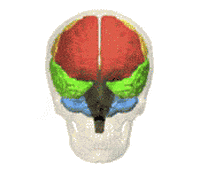
Exercise: Study the animation above, noting which colors represent which lobes or brain parts, then take the quiz.
Match lobes and structures with their corresponding colors by dragging the color to the correct box.
We recommend you view these movies in full screen mode. As before, watch for the extra information and quizzes!
Dr. Claudia Krebs, from the University of British Columbia, provides a nice introduction to the CNS in this video.
In this video, Dr. Suzanne Stensaas from the University of Utah, provides another good opportunity to view fixed brains and spinal cord, and important information about functional localization. Pay attention to the central and the pre- and post-central gyri.
Check your learning!
Drag the label to the correct leader or cortical site.
Of course, unless you are heading into neurosurgery or pathology, cadaver brains serve mostly as an aid to build your understanding of the 3D structure that will help you navigate clinical images that will be a large part of your professional life. Upcoming labs will be focusing on this, but here is a taste of how your knowledge should transfer. Use the slider to move between specimen and clinical views, and labeled and unlabeled views.
Cortical Localization
In this video clip, Dr. Stensaas guides us through the functional organization of the cortex. Note especially the location of the primary motor and somatosensory cortex, on either side of the central sulcus, and the language areas in the left hemisphere.
Watch for the interactive exercises and extra information!
Practice recognizing major landmarks and the functional areas related to them in the next 3 exercises. Move the sliders to first try to guess, then see the answers. A couple of functional areas are not on the cortical surface and are seen only in sections–these are revealed in the 3rd exercise.
Try this quick 7 question quiz on cortical functional localization.
Cerebellum
The cerebellum “little brain” contains millions of neurons and functionally has roles in balance, programming and coordination of motor activity.
Watch this brief introduction to the cerebellum by Dr. Stensaas.
Click on the check marks for more anatomical information.
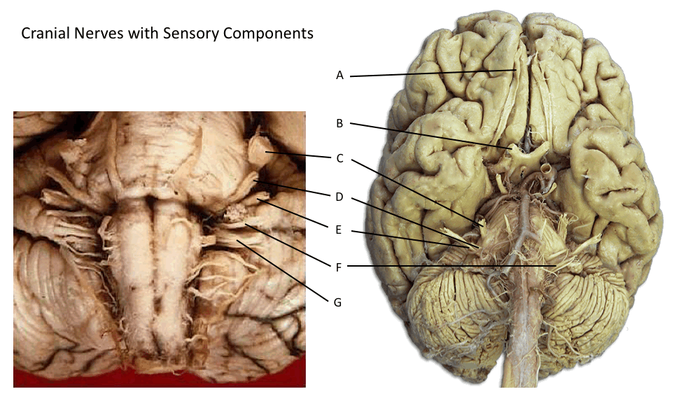
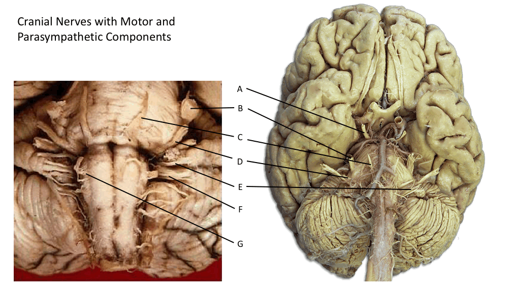
Brainstem Parts and Cranial Nerves
Dr. Krebs provides an overview of the parts of the brainstem in anterior, posterior and midsagittal views.
Check your learning!
In this video, Dr. Krebs demonstrates the attachment points of the cranial nerves.
Cranial nerve attachment points: Check your learning!
Here is a summary of cranial nerves for reference. You will be responsible for knowing cranial nerve exit foramina in upcoming labs, but that information is included here as you have already covered some of the information in other blocks (and I am a bit compulsive).
Sensory function of cranial nerves: check your learning by dragging and dropping the correct cranial nerve to the description of its function, referring to the image immediately below.
Motor and parasympathetic function of cranial nerves: check your learning by dragging and dropping the correct cranial nerve to the description of its function, referring to the image immediately below.
Spinal Cord
Dr. Claudia Krebs introduces the gross morphology of the human spinal cord.
Dr. Suzanne Stensaas surveys regional variations in the gray and white matter of the spinal cord and describes a simple monosynaptic reflex.
Be sure you can distinguish dorsal (posterior) from ventral (anterior) cord and rootlets. These images will help.
Check your learning!