16 Bundle Branch Blocks, a review
Bundle branch blocks represent a delayed conduction in the His Purkinje system. If there is a characteristic ECG pattern of this delay, we can define right and left bundle branch blocks. Remember, the direction of current is seen from all angles simultaneously (V1 is opposite V6 on the chest which is why the the voltage deflection is almost always opposite). It is best to use V1 and V6 when evaluating for bundle branch blocks when the QRS duration is prolonged.
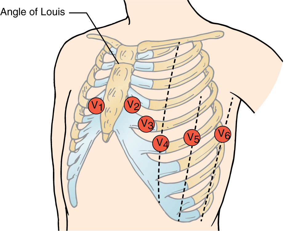
Here we see the proper placement of the precordial (chest) leads.
Here is a reminder of the key electrical components of the heart. Notice the His bundle promptly splits into the left and right bundles. If there is a ischemic (or other) injury to this electrical tissue, conduction will not go down the fast bundles. Conduction will then occur slowly via cell-to-cell connections (gap junctions). This delay in time is seen as width on the ECG. 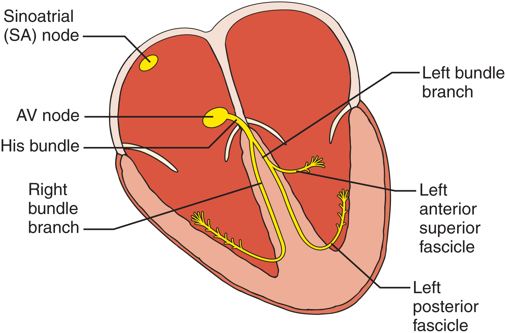
Here we see a drawing of the ‘wave’ of conduction through the ventricles in brown in right bundle branch block. The first signal is rightward via the left bundle [negative in V6 but positive in V1], then proceeds quickly via the left bundle to the LV [negative in V1 and positive in V6]. The slow cell-to-cell duction then proceeds rightward (positive in V1 and negative in V6). 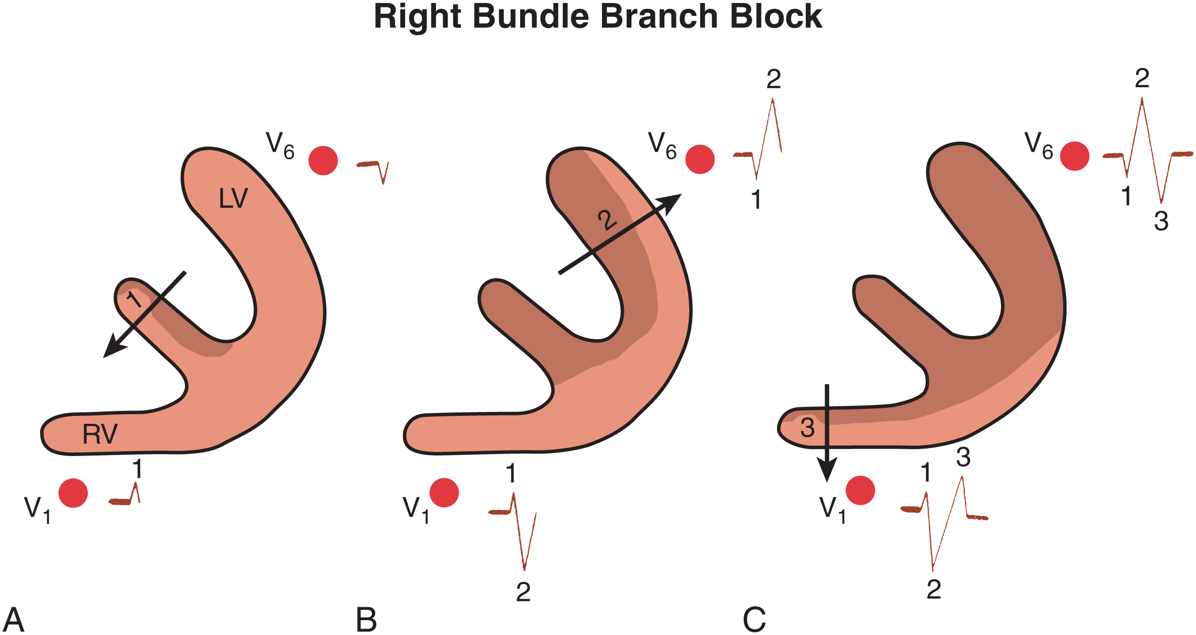
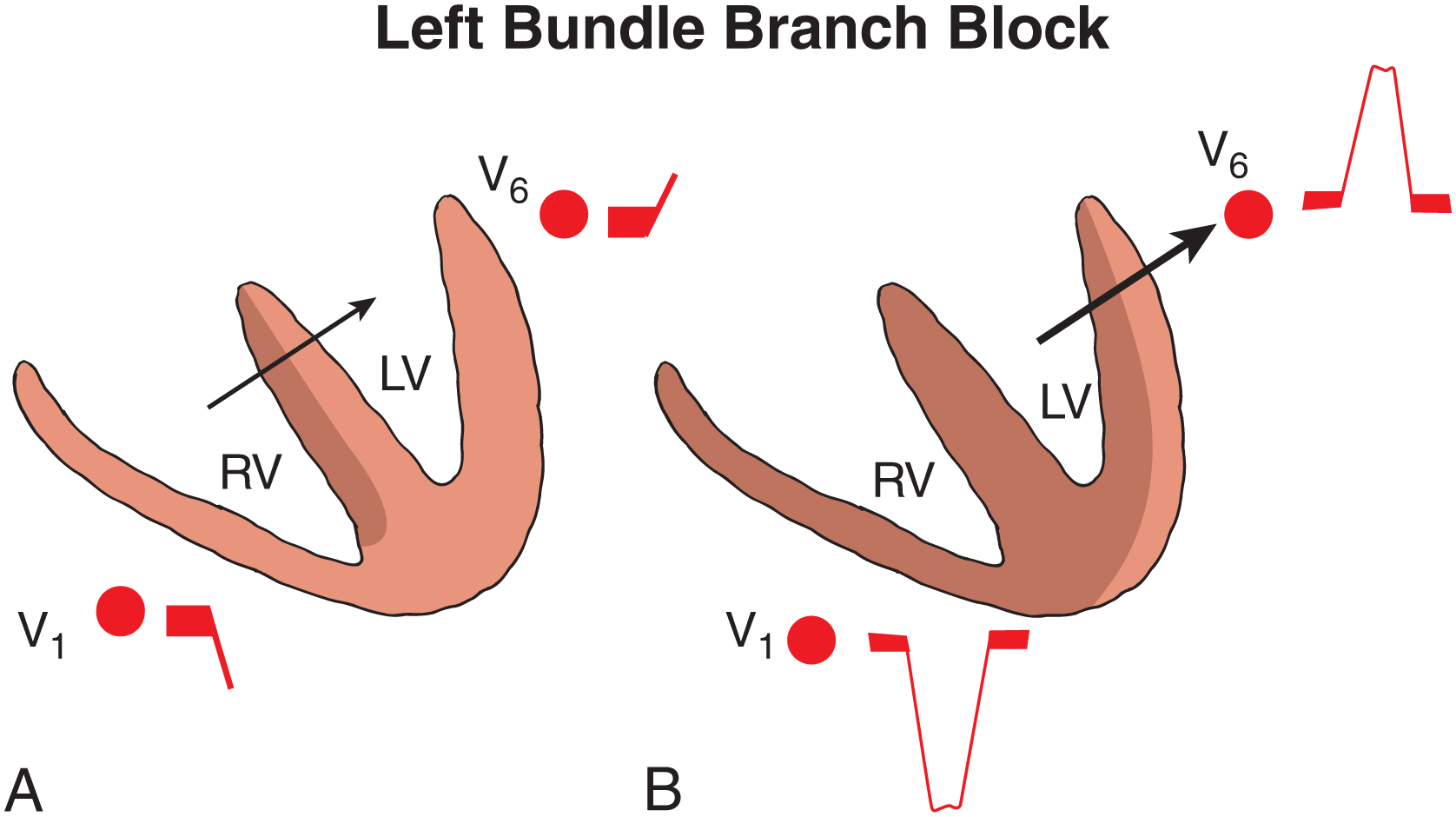
Here we see a drawing of the ‘wave’ of conduction through the ventricles in brown in left bundle branch block. The signal is leftward starting at the septum via the right bundle depolarizing some of the septum and RV [positive in V6 and negative in V1]. The slow cell-to-cell duction proceeds leftward the whole time all the way to the lateral wall which is why the entire QRS is positive in V6 and negative in V1. As the right ventricle is much smaller the left, RV conduction is not seen.
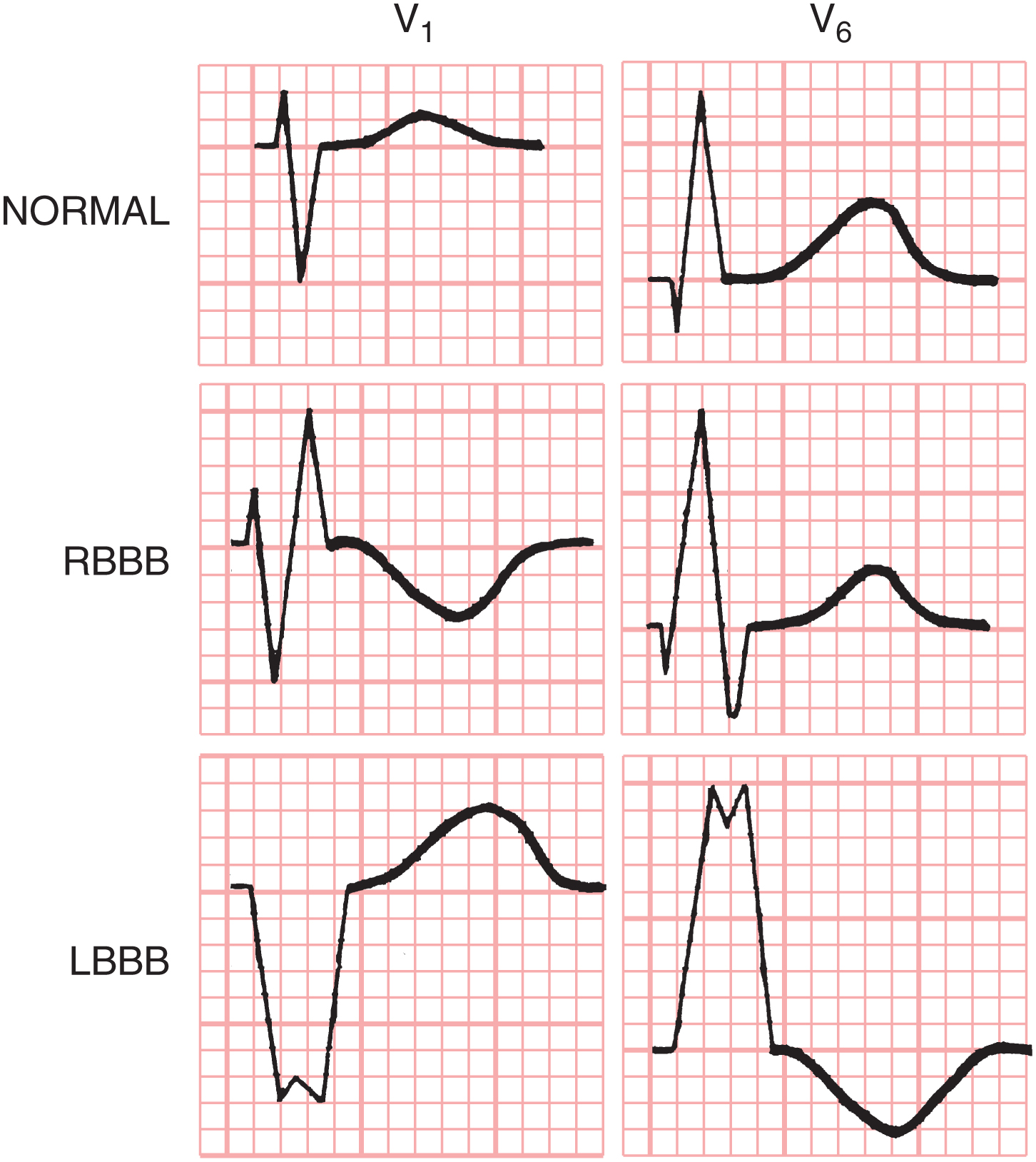
Here is a quick schematic to think about normal QRS morphology, along with left and right bundle branch blocks. Of course, this is focusing on leads V1 and V6.